Copyright
©The Author(s) 2021.
World J Clin Cases. Jun 6, 2021; 9(16): 4040-4045
Published online Jun 6, 2021. doi: 10.12998/wjcc.v9.i16.4040
Published online Jun 6, 2021. doi: 10.12998/wjcc.v9.i16.4040
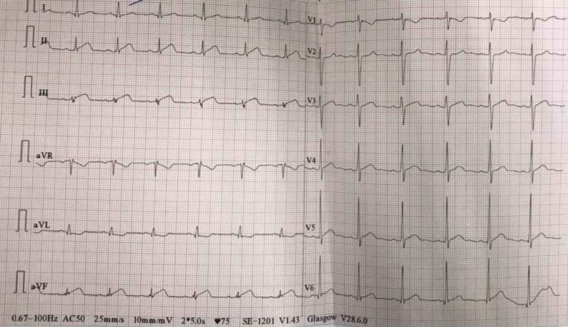
Figure 1
Electrocardiogram with sinus rhythm and ST-segment elevation in DII, DIII and aVF leads.
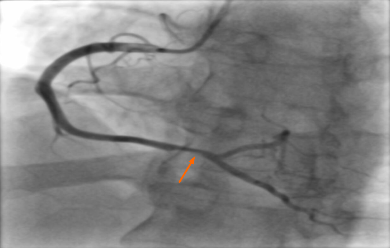
Figure 2
Right coronary artery showing mild stenotic lesion with thrombus at crux cordis (arrow).
- Citation: Scafa-Udriste A, Popa-Fotea NM, Bataila V, Calmac L, Dorobantu M. Acute inferior myocardial infarction in a young man with testicular seminoma: A case report. World J Clin Cases 2021; 9(16): 4040-4045
- URL: https://www.wjgnet.com/2307-8960/full/v9/i16/4040.htm
- DOI: https://dx.doi.org/10.12998/wjcc.v9.i16.4040


