Copyright
©The Author(s) 2021.
World J Clin Cases. Jun 6, 2021; 9(16): 3996-4000
Published online Jun 6, 2021. doi: 10.12998/wjcc.v9.i16.3996
Published online Jun 6, 2021. doi: 10.12998/wjcc.v9.i16.3996
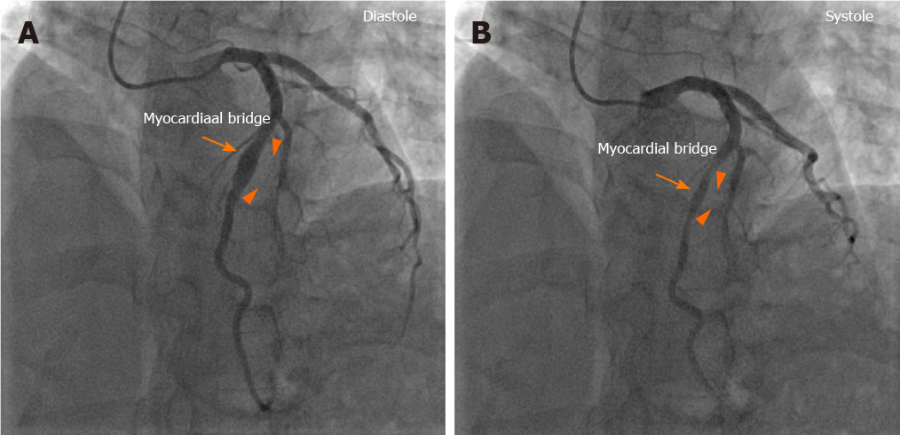
Figure 1 Images of coronary angiography.
A: Coronary angiography showed the coronary artery aneurysm (CAA) in the middle left anterior descending artery during diastole; B: Coronary angiography showed that the CAA was compressed by the myocardial bridge during systole. The arrow indicates the location of the myocardial bridge, and the triangles indicate the start and end of the CAA.
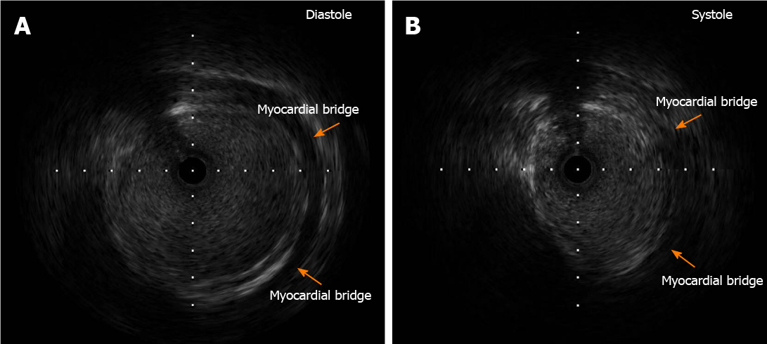
Figure 2 Images of intravascular ultrasound.
A: Intravascular ultrasound showed the coronary artery aneurysm and myocardial bridge during diastole; B: Intravascular ultrasound showed that the coronary artery aneurysm was compressed by the myocardial bridge during systole. The arrows indicate the location of the myocardial bridge.
- Citation: Ye Z, Dong XF, Yan YM, Luo YK. Coronary artery aneurysm combined with myocardial bridge: A case report. World J Clin Cases 2021; 9(16): 3996-4000
- URL: https://www.wjgnet.com/2307-8960/full/v9/i16/3996.htm
- DOI: https://dx.doi.org/10.12998/wjcc.v9.i16.3996


