Copyright
©The Author(s) 2021.
World J Clin Cases. Jun 6, 2021; 9(16): 3979-3987
Published online Jun 6, 2021. doi: 10.12998/wjcc.v9.i16.3979
Published online Jun 6, 2021. doi: 10.12998/wjcc.v9.i16.3979
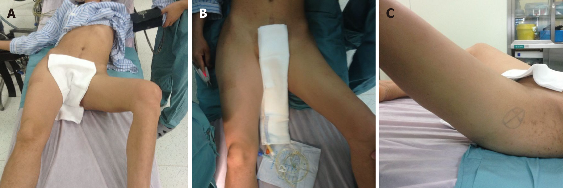
Figure 1 Physical examination.
A: Front view of the patient showing that there was compensatory scoliosis towards the left, and the pelvis was lower on the left side; B and C: Front and lateral view of the left hip showing that the hip was fixed in 40° of flexion, 45° of abduction, and 30° of external rotation.
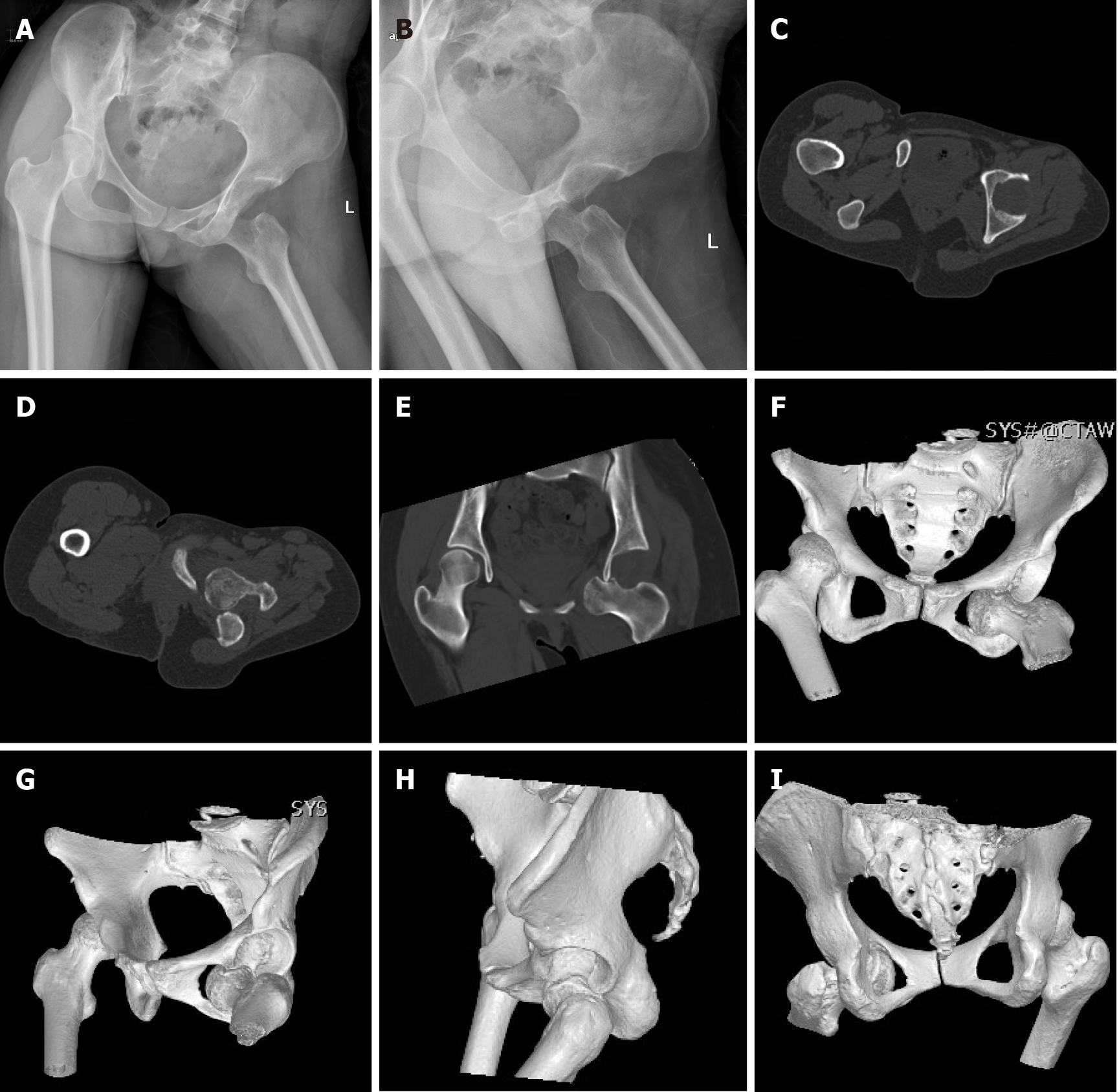
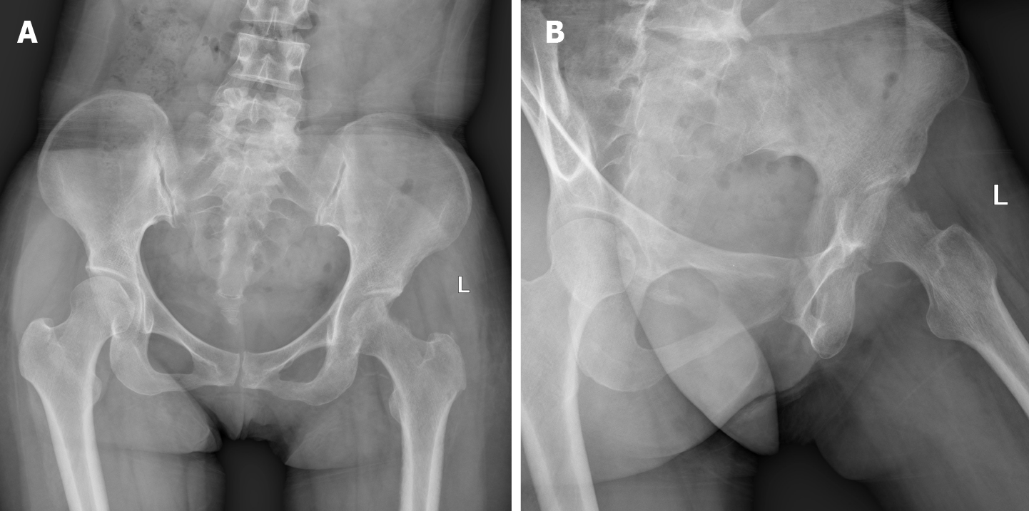
Figure 2 Preoperative plain radiographs and computed tomography scans.
A and B: Plain radiographs at the first examination showing obturator dislocation of the left hip; C-I: Axial, coronal, and three-dimensional reconstructed computed tomography images showing that the femoral head lay anteriorly and inferiorly to the obturator foramen, with impaction fracture at the superolateral aspect of the left femoral head without associated fracture of the acetabulum.
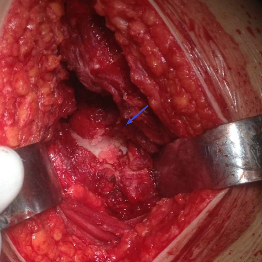
Figure 3 Intraoperative finding.
The anterosuperior aspect of the femoral head (blue arrow) showed collapse and articular damage.
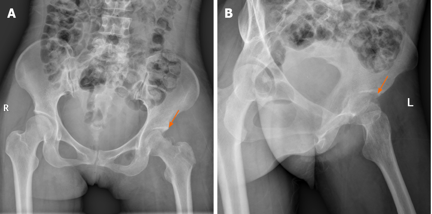
Figure 4 Postoperative plain radiographs.
A: Anteroposterior (orange arrow); B: Lateral plain radiographs of the left hip showed concentric alignment of the left hip and impaction fracture (orange arrow) of the femoral head.
Figure 5 Plain radiographs at 3 mo after surgery.
A: Anteroposterior; B: Lateral plain radiographs of the left hip showed that the left hip was homocentric, and there was no sign of posttraumatic arthritis or avascular necrosis.
- Citation: Li WZ, Wang JJ, Ni JD, Song DY, Ding ML, Huang J, He GX. Old unreduced obturator dislocation of the hip: A case report. World J Clin Cases 2021; 9(16): 3979-3987
- URL: https://www.wjgnet.com/2307-8960/full/v9/i16/3979.htm
- DOI: https://dx.doi.org/10.12998/wjcc.v9.i16.3979


