Copyright
©The Author(s) 2021.
World J Clin Cases. Jun 6, 2021; 9(16): 3960-3965
Published online Jun 6, 2021. doi: 10.12998/wjcc.v9.i16.3960
Published online Jun 6, 2021. doi: 10.12998/wjcc.v9.i16.3960
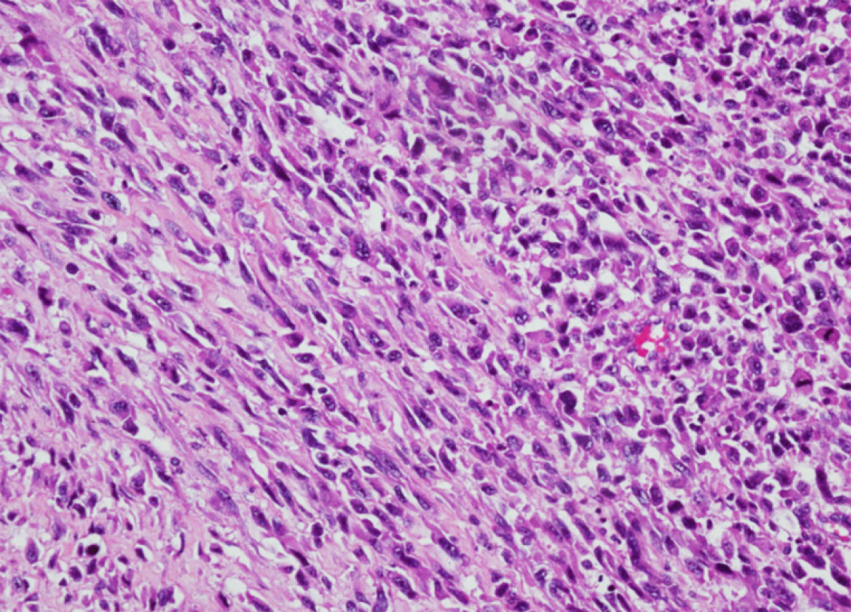
Figure 1 The undifferentiated intimal sarcoma is composed of spindle and pleomorphic cells with obvious atypia.
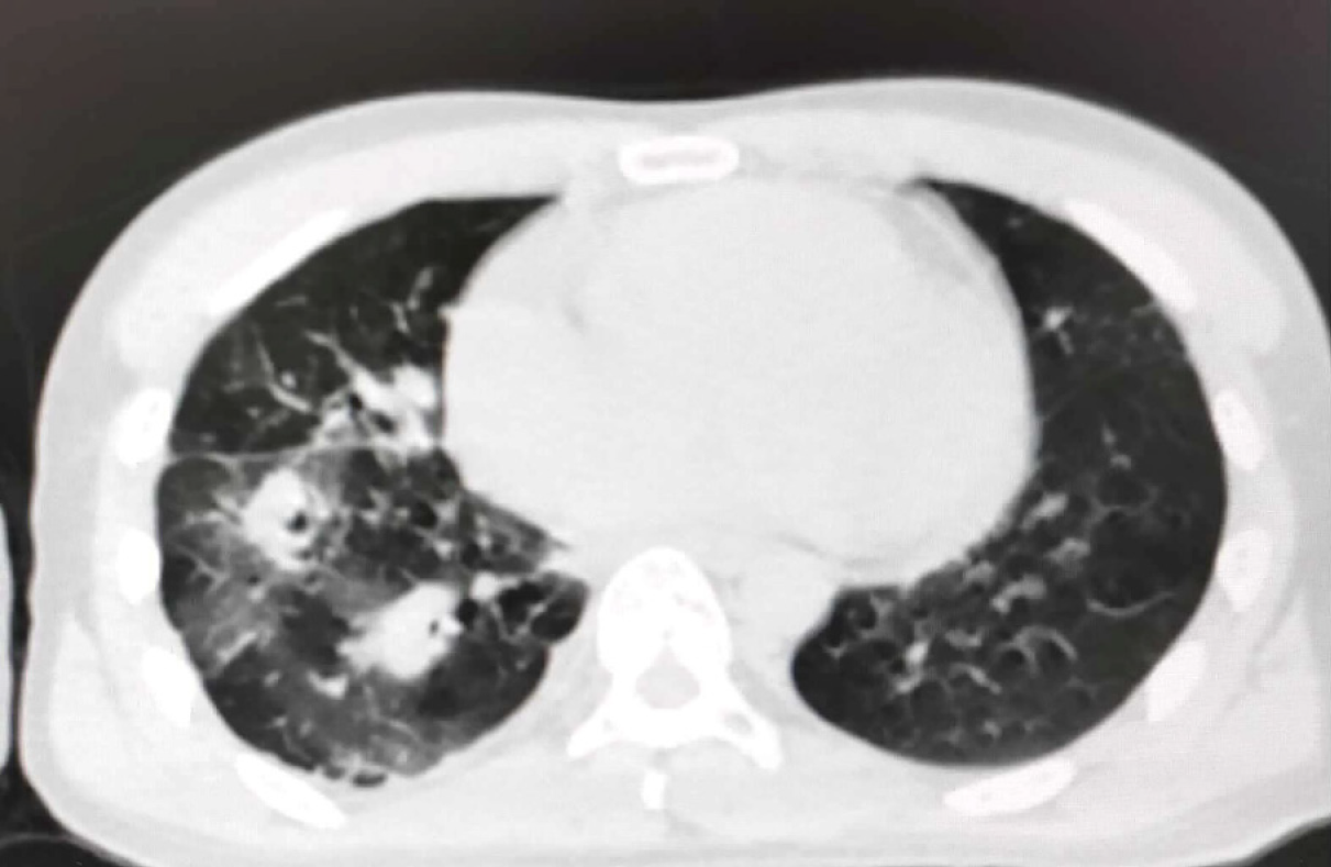
Figure 2 Computed tomography images revealing multiple nodules and patchy images in the right lung.
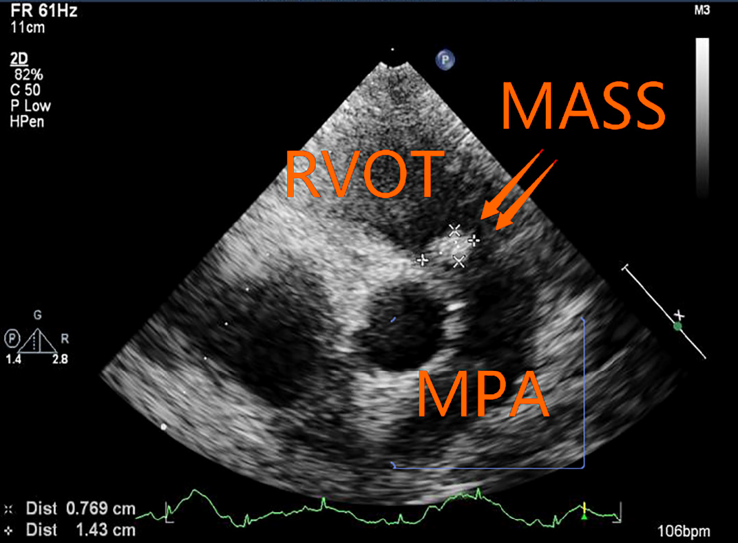
Figure 3 A solid mass of 14 mm × 7 mm was detected in the pulmonary artery.
RVOT: Right ventricular outflow tract; MPA: Main pulmonary artery.
- Citation: Li X, Hong L, Huo XY. Undifferentiated intimal sarcoma of the pulmonary artery: A case report. World J Clin Cases 2021; 9(16): 3960-3965
- URL: https://www.wjgnet.com/2307-8960/full/v9/i16/3960.htm
- DOI: https://dx.doi.org/10.12998/wjcc.v9.i16.3960


