Copyright
©The Author(s) 2021.
World J Clin Cases. Jun 6, 2021; 9(16): 3943-3950
Published online Jun 6, 2021. doi: 10.12998/wjcc.v9.i16.3943
Published online Jun 6, 2021. doi: 10.12998/wjcc.v9.i16.3943
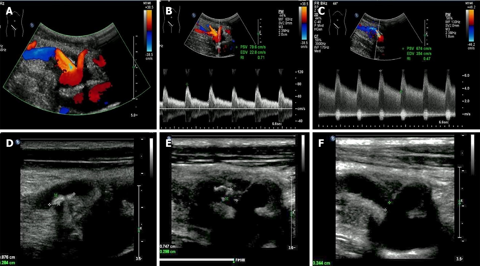
Figure 1 Ultrasonographic examination.
A: Two artery trunks anastomosed with the side of the external iliac artery were detected; B: Peak systolic velocity (PSV) of the low pole artery was 79.6 cm/s; C: PSV of 674 cm/s with color aliasing at the proximal site of the main renal trunk was detected; D and E: Multiple hypo-echoic plaques attached to the wall of the main renal artery trunk was detected; F: The residual lumen at the proximal site of the main renal artery trunk was 0.2 cm in diameter.
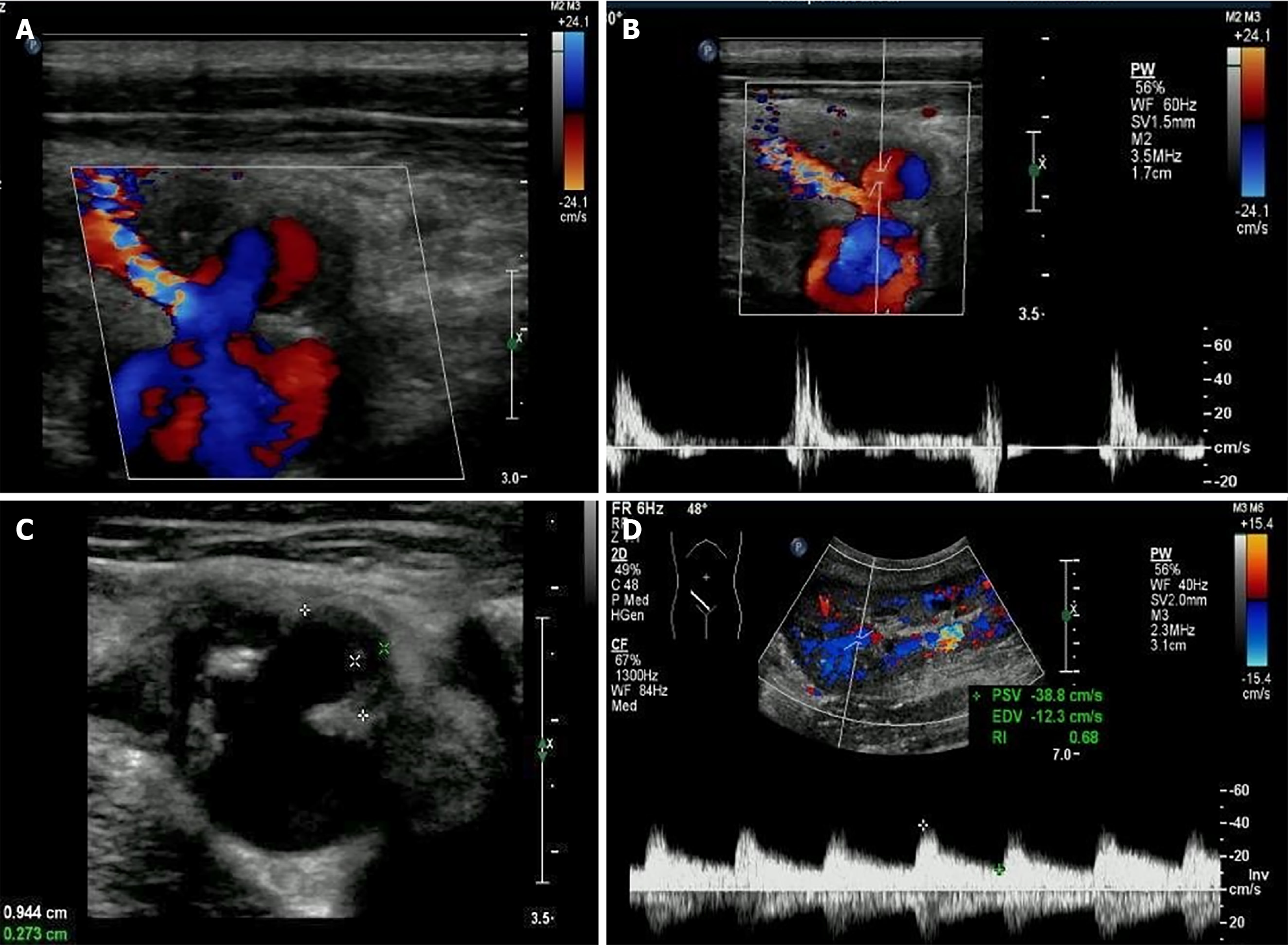
Figure 2 Extra-renal pseudo-aneurysm under ultrasonography.
A: Blood flow signal showing as swirling pattern was visible; B: To-and-fro disorganized arterial spectrum was detected with pulsed-wave Doppler; C: Hypo-echoic thrombus with thickness of 0.3 cm was observed inside the cystic structure; D: Prolonged acceleration time was longer than 70 ms of the related area inside the graft.
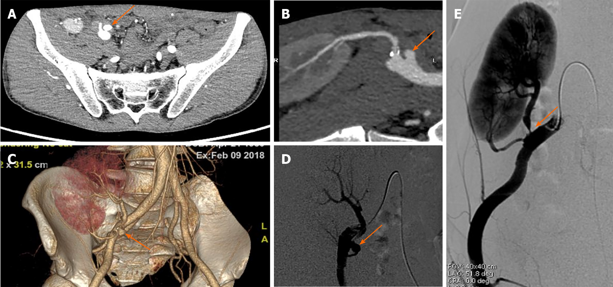
Figure 3 Computed tomography angiography and digital subtraction angiography manifestations of the extra-renal pseudo-aneurysm.
A-C: Extra-renal pseudo-aneurysm (EPSA at the anastomotic site was seen by computed tomography angiography examination (orange arrows); D and E: Severe stenosis at the proximal site of the main renal artery trunk (E) with EPSA at the anastomosis with the external iliac artery (D) was confirmed by digital subtraction angiography examination (orange arrows).
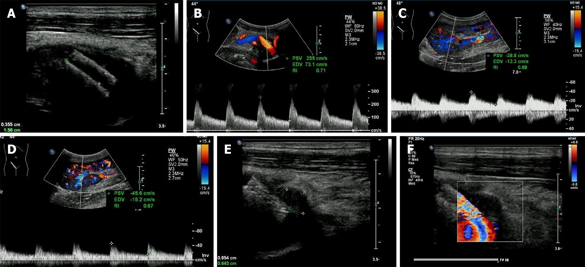
Figure 4 Ultrasonographic follow-ups post-treatment.
A and B: Stent was detected at the proximal site the of the main renal artery trunk and the peak systolic velocity decreased to 255 cm/s after stent implantation; C and D: The prolonged acceleration time disappeared (D) compared with pre-treatment manifestation (C); E and F: The extra-renal pseudo-aneurysm was completely filled with hypo-echoic thrombosis 8 mo after stenting.
- Citation: Xu RF, He EH, Yi ZX, Li L, Lin J, Qian LX. Diagnosis and spontaneous healing of asymptomatic renal allograft extra-renal pseudo-aneurysm: A case report. World J Clin Cases 2021; 9(16): 3943-3950
- URL: https://www.wjgnet.com/2307-8960/full/v9/i16/3943.htm
- DOI: https://dx.doi.org/10.12998/wjcc.v9.i16.3943


