Copyright
©The Author(s) 2021.
World J Clin Cases. Jun 6, 2021; 9(16): 3919-3926
Published online Jun 6, 2021. doi: 10.12998/wjcc.v9.i16.3919
Published online Jun 6, 2021. doi: 10.12998/wjcc.v9.i16.3919
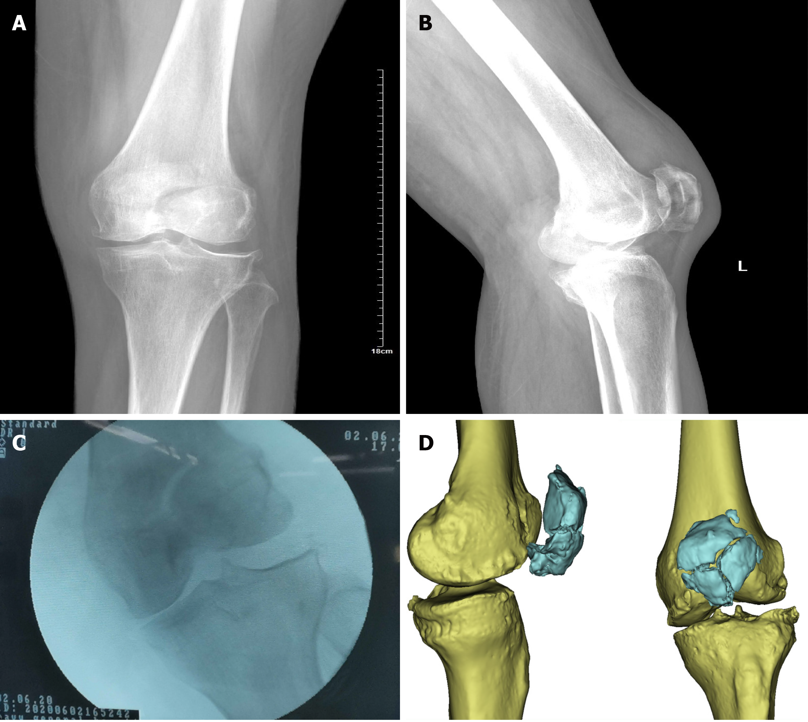
Figure 1 Preoperative images.
A: Anteroposterior view; B: Lateral view; C: Varus stress radiography of the left knee showing the patella fracture and classic anteromedial osteoarthritis; D: Preoperative computed tomography of the left knee showing the comminuted patella fracture.
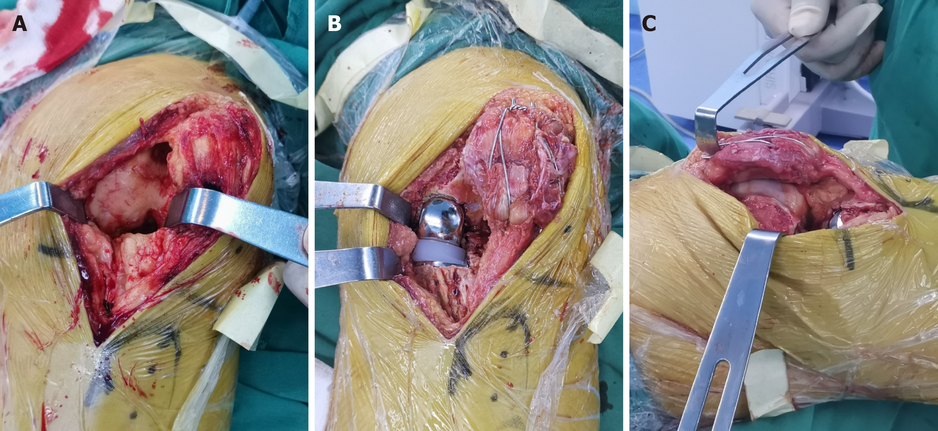
Figure 2 Intraoperative photographs.
A: The full layer cartilage of the distal part of the medial condyle of the femur and the anteromedial full-thickness cartilage of the medial tibial plateau were worn. The anterior cruciate ligament was intact and the cartilage of the lateral compartment on both sides was intact; B and C: Photographs after implanting the patellar internal fixations and Oxford prosthesis. There was no impingement between them.
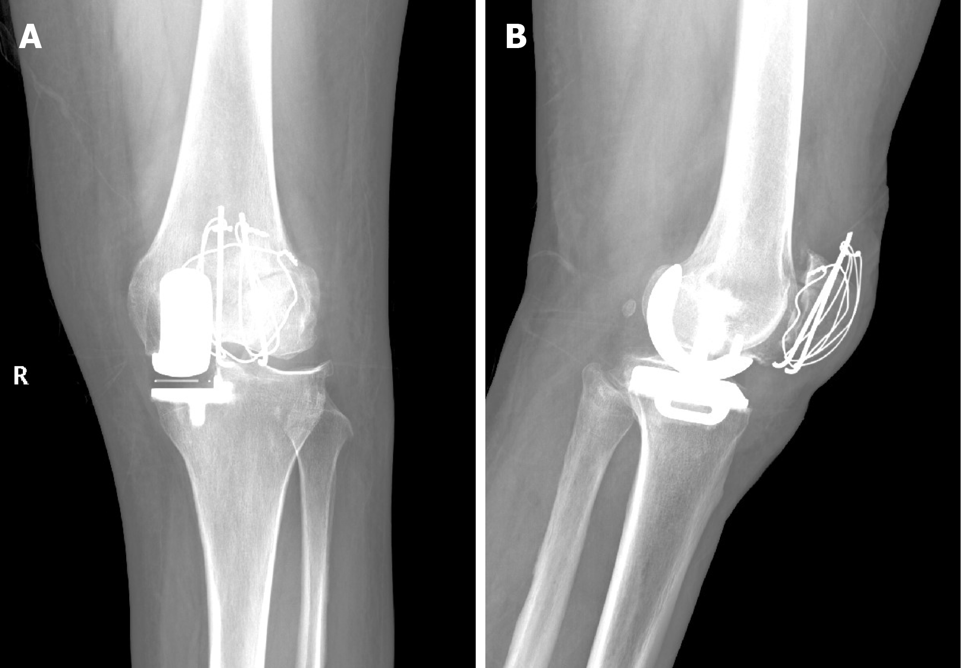
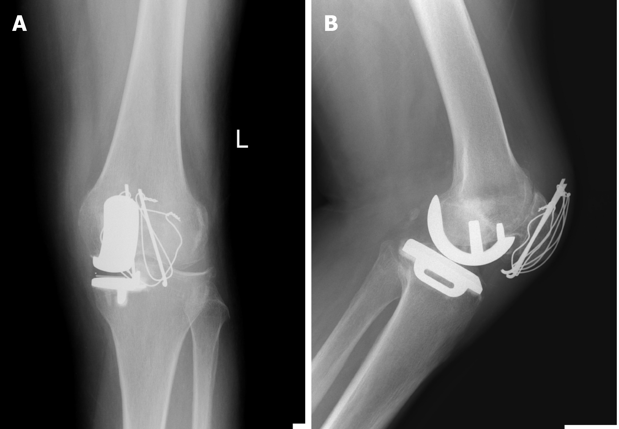
Figure 3 Postoperative radiographs obtained at 3 d after surgery showing good reduction of the patellar fracture and an appropriate Oxford unicompartmental knee arthroplasty.
A: Anteroposterior view; B: Lateral view.
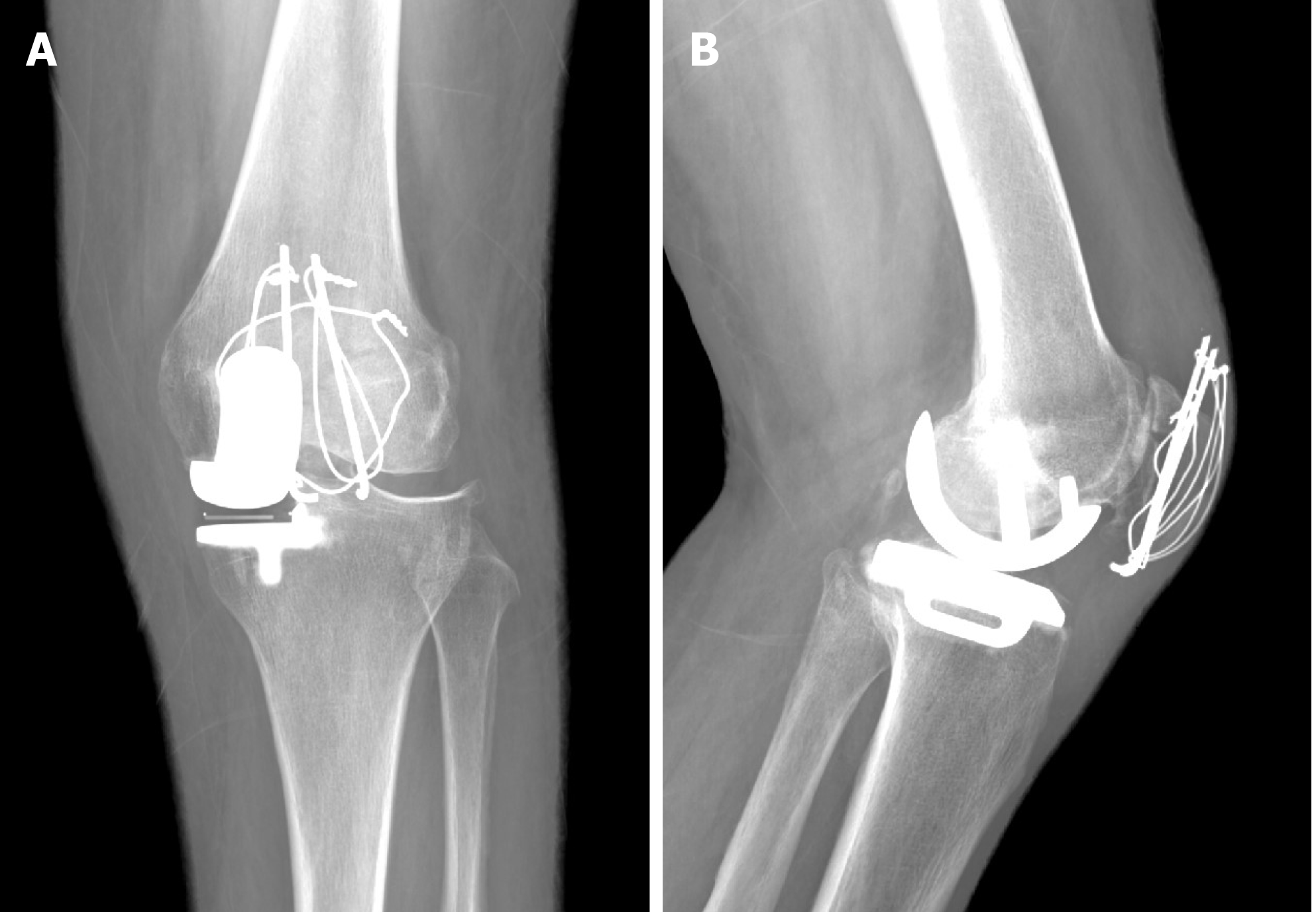
Figure 4 Postoperative radiographs obtained at 8 wk after surgery.
A: Anteroposterior view; B: Lateral view.
Figure 5 Postoperative radiographs obtained at 12 wk after surgery.
A: Anteroposterior view; B: Lateral view.
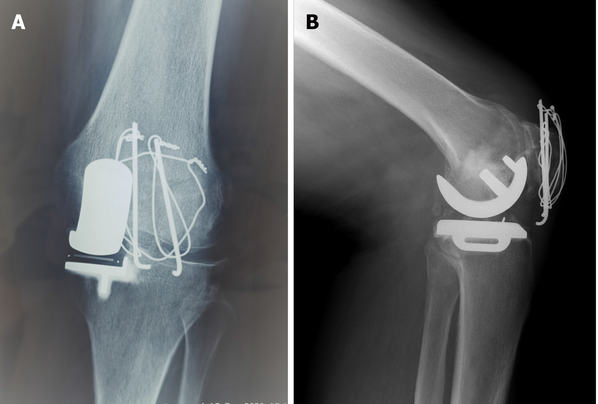
Figure 6 Postoperative radiographs obtained 20 wk after surgery.
A: Anteroposterior view; B: Lateral view.
- Citation: Nan SK, Li HF, Zhang D, Lin JN, Hou LS. Internal fixation and unicompartmental knee arthroplasty for an elderly patient with patellar fracture and anteromedial osteoarthritis: A case report. World J Clin Cases 2021; 9(16): 3919-3926
- URL: https://www.wjgnet.com/2307-8960/full/v9/i16/3919.htm
- DOI: https://dx.doi.org/10.12998/wjcc.v9.i16.3919


