Copyright
©The Author(s) 2021.
World J Clin Cases. Jun 6, 2021; 9(16): 3914-3918
Published online Jun 6, 2021. doi: 10.12998/wjcc.v9.i16.3914
Published online Jun 6, 2021. doi: 10.12998/wjcc.v9.i16.3914
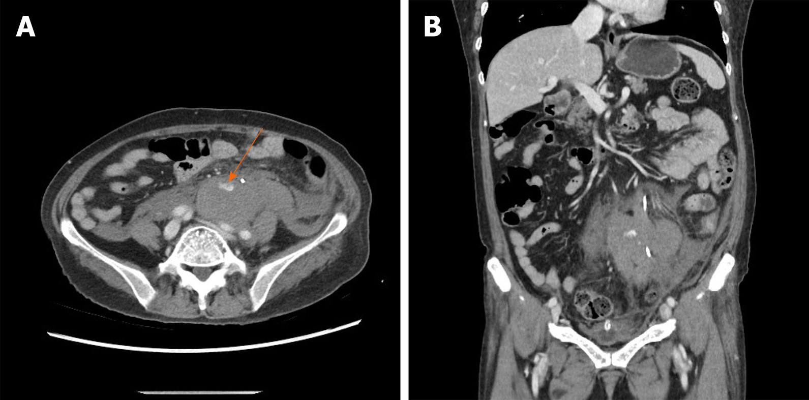
Figure 1 Computed tomography scans showing a periureteral hematoma in transverse view and coronal view.
A: Transverse view computed tomography scan. The orange arrow marks the extravasation of contrast medium; B: Coronal view computed tomography scan.
- Citation: Choi T, Choi J, Min GE, Lee DG. Massive retroperitoneal hematoma as an acute complication of retrograde intrarenal surgery: A case report. World J Clin Cases 2021; 9(16): 3914-3918
- URL: https://www.wjgnet.com/2307-8960/full/v9/i16/3914.htm
- DOI: https://dx.doi.org/10.12998/wjcc.v9.i16.3914


