Copyright
©The Author(s) 2021.
World J Clin Cases. Jun 6, 2021; 9(16): 3848-3857
Published online Jun 6, 2021. doi: 10.12998/wjcc.v9.i16.3848
Published online Jun 6, 2021. doi: 10.12998/wjcc.v9.i16.3848
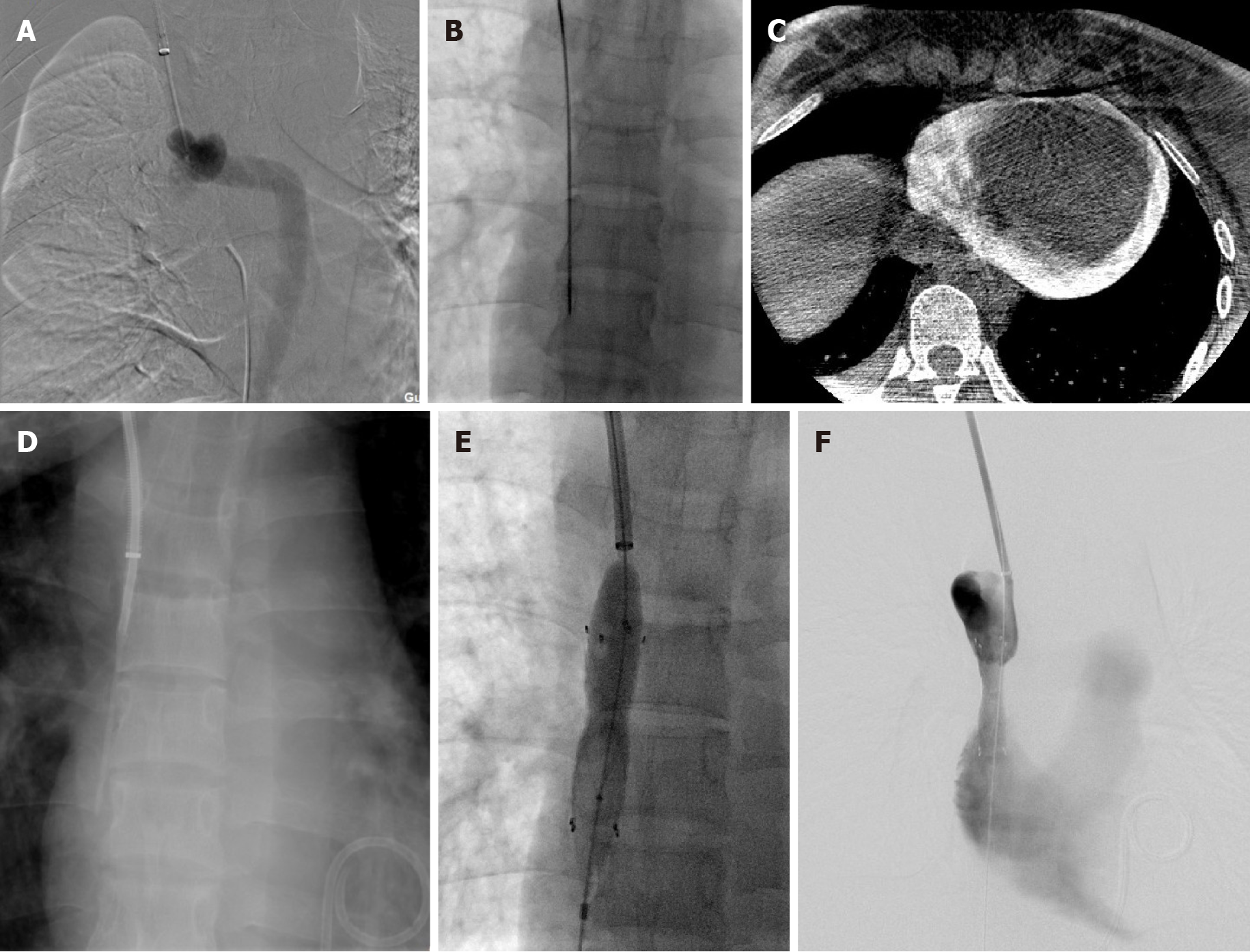
Figure 1 A 25-yr-old female with a sharp recanalization procedure due to superior vena cava obstruction.
A: Before sharp recanalization, angiography showed superior vena cava occlusion; B and C: After puncturing the occluded segment (B), the patient complicated with acute pericardial tamponade (C); D: Sharp recanalization succeeded at the second surgery; E: Percutaneous transluminal angioplasty and percutaneous transluminal stenting were performed; F: Venous blood of the superior vena cava.
Figure 2 A 25-yr-old female.
Postoperative review of sharp recanalization procedure for superior vena cava obstruction. A and B: Coronal (A) and sagittal (B) computed tomography of superior vena cava; C: Digital subtraction angiography of superior vena cava.
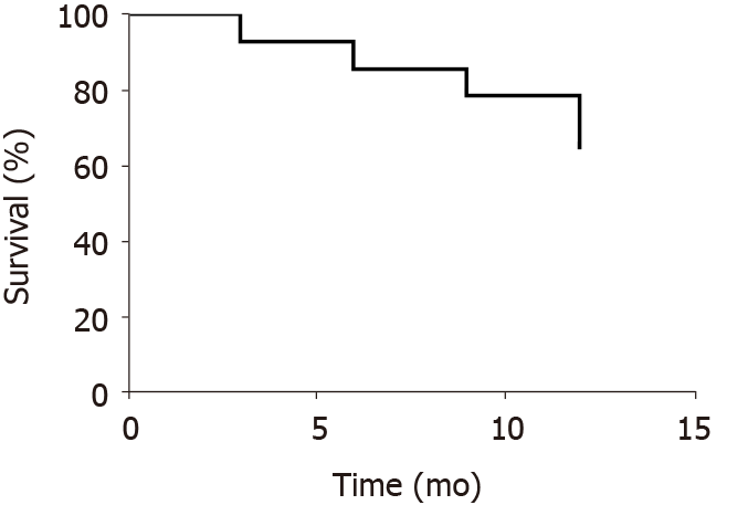
Figure 3 Superior vena cava first patency rate after sharp recanalization surgery.
- Citation: Wu XW, Zhao XY, Li X, Li JX, Liu ZY, Huang Z, Zhang L, Sima CY, Huang Y, Chen L, Zhou S. Effectiveness of sharp recanalization of superior vena cava-right atrium junction occlusion. World J Clin Cases 2021; 9(16): 3848-3857
- URL: https://www.wjgnet.com/2307-8960/full/v9/i16/3848.htm
- DOI: https://dx.doi.org/10.12998/wjcc.v9.i16.3848


