Copyright
©The Author(s) 2021.
World J Clin Cases. May 26, 2021; 9(15): 3741-3751
Published online May 26, 2021. doi: 10.12998/wjcc.v9.i15.3741
Published online May 26, 2021. doi: 10.12998/wjcc.v9.i15.3741
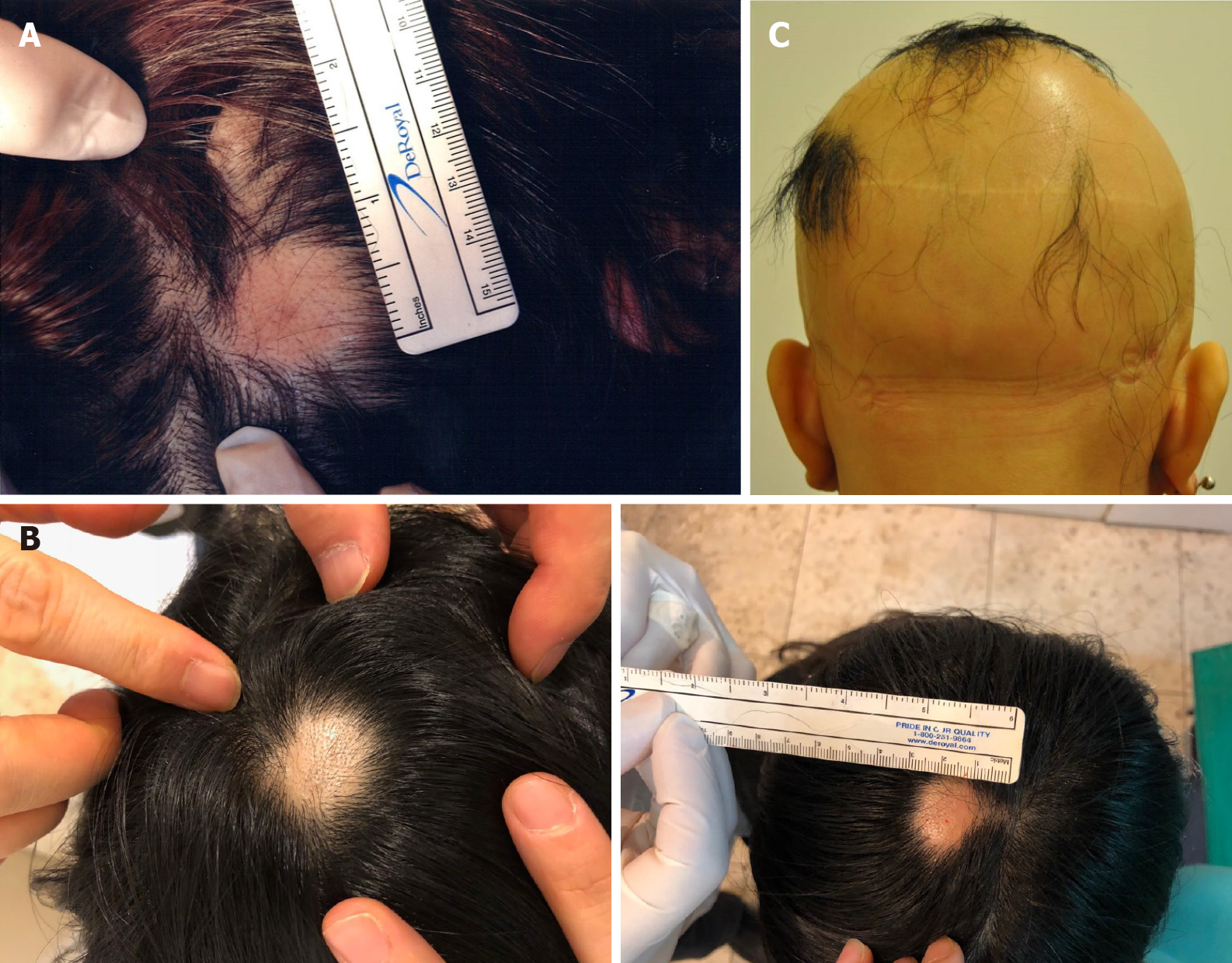
Figure 1 Alopecia areata sites of the patients before treatment.
A: The image represents the alopecia lesion sites of case 1 before transplantation of minimally manipulated umbilical cord-derived mesenchymal stem cells; B: Case 2 before transplantation of minimally manipulated umbilical cord-derived mesenchymal stem cells; C: Case 3 before transplantation of minimally manipulated umbilical cord-derived mesenchymal stem cells.
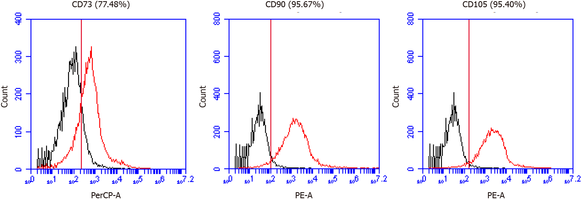
Figure 2 Mesenchymal stem cell marker expression in minimally manipulated umbilical cord-derived mesenchymal stem cells.
A: The expression marker tested was CD73 (77.48%); B: CD90 (95.67%); C: CD105 (95.40%).
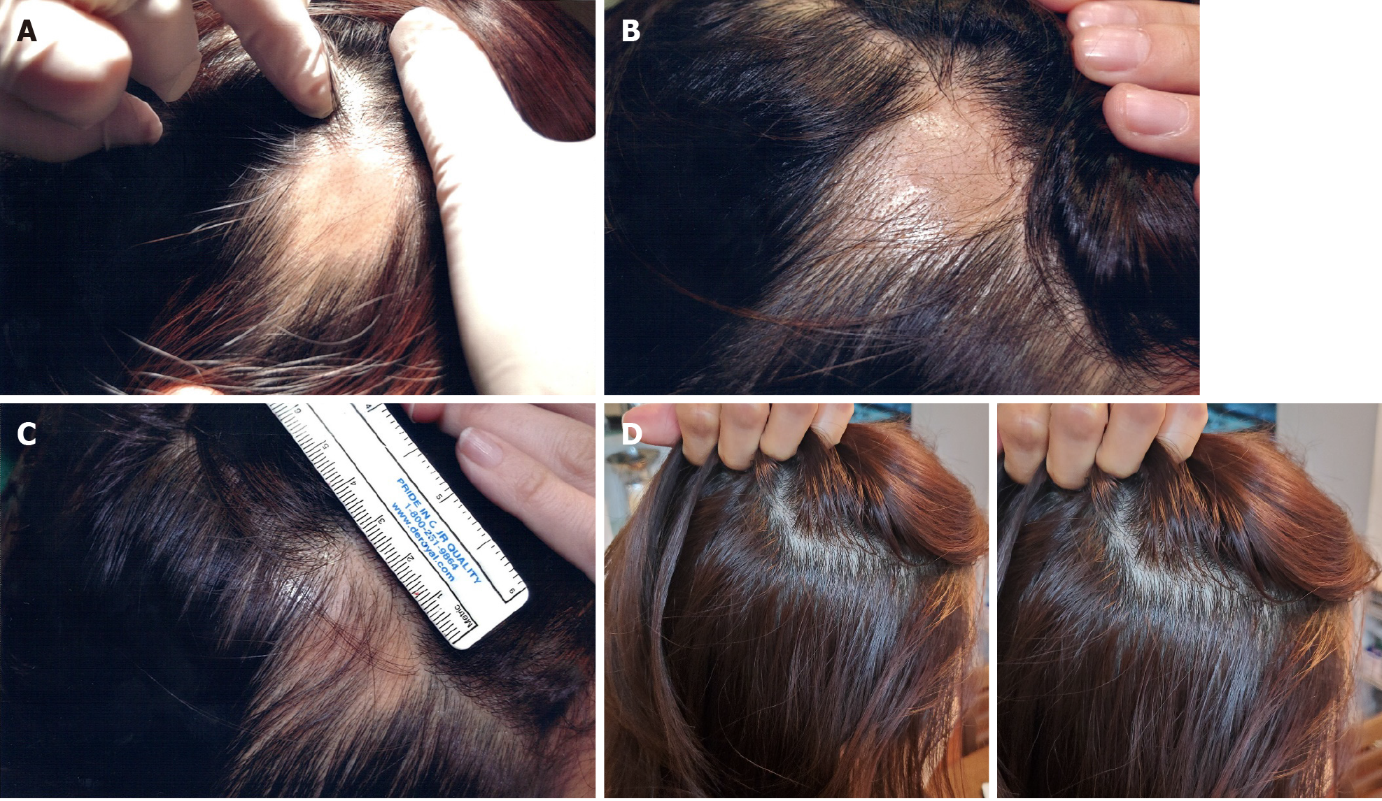
Figure 3 Visible changes at the lesion sites of case 1 during and after treatment.
A and B: There were no significant changes observed at (A) day 33 or (B) day 53 after the first transplant; C: Hair covered the lesion site at day 164 after the first transplant; D: These images were taken 22 mo after the final transplant. The patient was completely cured and maintained the hair growth. The images represent the lesion on the right side of the scalp.
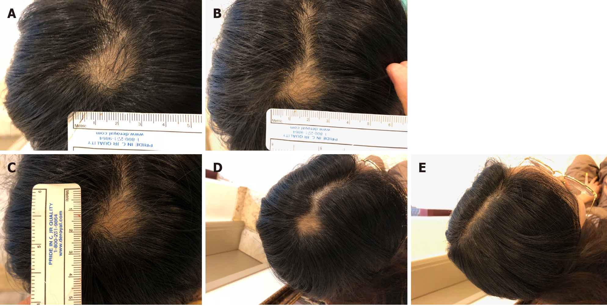
Figure 4 Visible changes at the lesion site of case 2 during and after treatment.
A: Images of the lesion site were obtained at day 26 after the first transplant; B: Day 43 after the first transplant; C: Day 58 after the first transplant; D: Day 89 after the first transplant; E: Day 117 after the first transplant.
Figure 5 Visible changes during the treatment process of case 3.
A: The images show the back of the head at day 160 after the first transplant; B: Day 203 after the first transplant; C: Day 226 after the first transplant.
- Citation: Ahn H, Lee SY, Jung WJ, Lee KH. Alopecia treatment using minimally manipulated human umbilical cord-derived mesenchymal stem cells: Three case reports and review of literature. World J Clin Cases 2021; 9(15): 3741-3751
- URL: https://www.wjgnet.com/2307-8960/full/v9/i15/3741.htm
- DOI: https://dx.doi.org/10.12998/wjcc.v9.i15.3741


