Copyright
©The Author(s) 2021.
World J Clin Cases. May 26, 2021; 9(15): 3704-3710
Published online May 26, 2021. doi: 10.12998/wjcc.v9.i15.3704
Published online May 26, 2021. doi: 10.12998/wjcc.v9.i15.3704
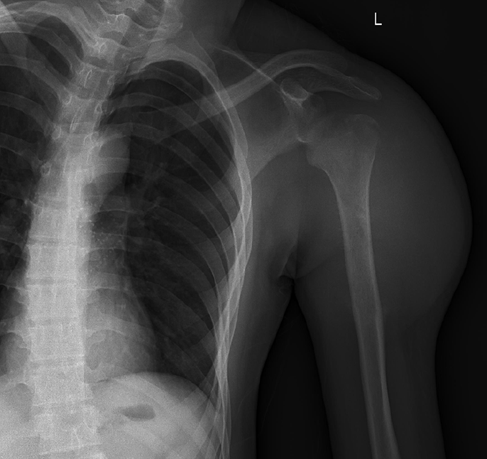
Figure 1 Posteroanterior shoulder joint radiograph.
A linear low-density shadow at the greater tuberosity of the left humerus and small flakes in the upper medullary cavity of the left humerus with slightly reduced density are seen.
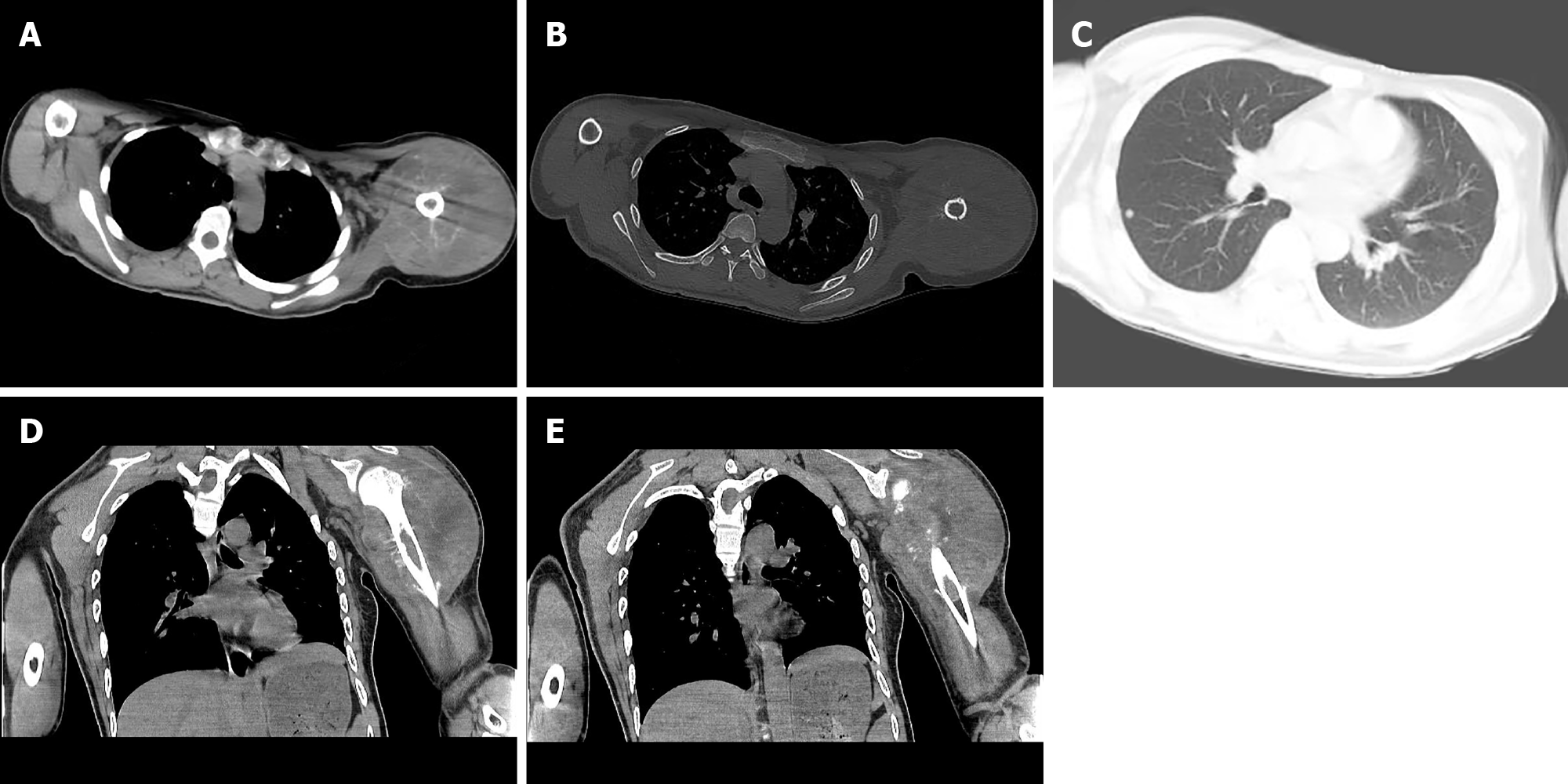
Figure 2 Computed tomography of left shoulder joint reveals a soft tissue mass of the left shoulder.
A: Strip-like calcifications in the mass; B: The adjacent bone shows an insect-like zone of destruction; C: Small, rounded solid nodules in the lower lobe of the right lung; D: The adjacent bone shows a needle-like periosteal reaction; E: Invasion of the marrow cavity.
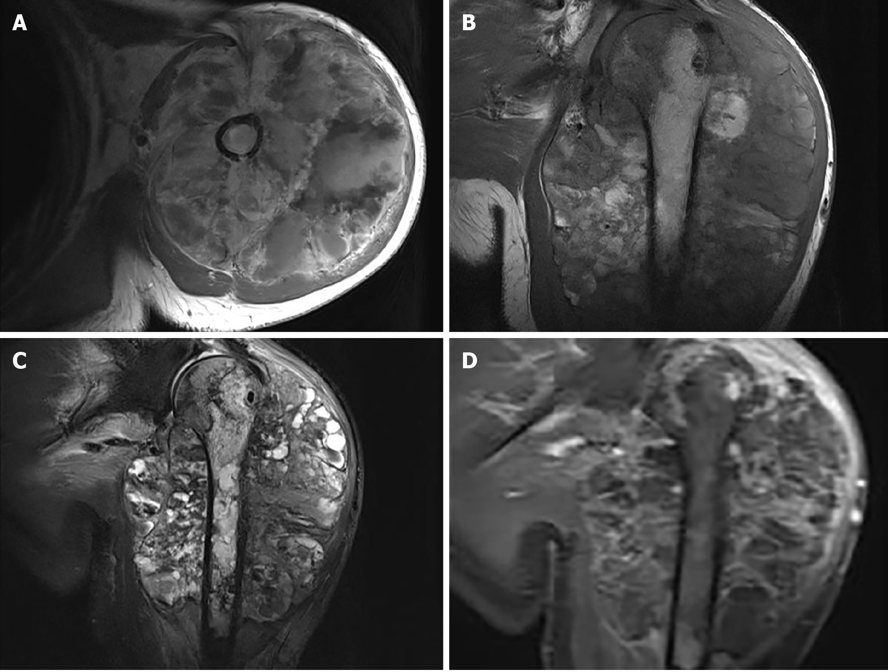
Figure 3 Upper arm magnetic resonance imaging.
A and B: Mixed moderate hypointensity and hyperintensity on axial (A) and coronal (B) T1-weighted images; C: mixed hyperintensity on coronal fat-saturated T2-weighted images; D: T1-weighted images with contrast demonstrate heterogeneous enhancement of the lesion.
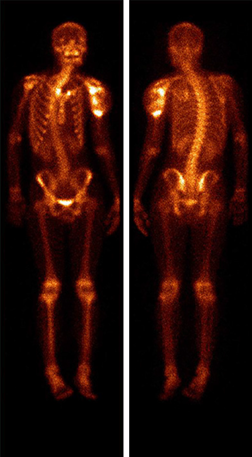
Figure 4 Whole-body 99mTc-methylene diphosphonate bone scan imaging.
Abnormal distribution of radioactive label concentration and increased activity are seen in the left shoulder joint and proximal humerus.
Figure 5 Histopathological images.
A: A giant cell tumor with polygonal mononuclear cells and multinucleated osteoclast-like giant cells (hematoxylin and eosin, × 200); B: Immunohistochemical staining revealed strong CD163 positivity (hematoxylin and eosin, × 200); C: Immunohistochemical staining revealed high CD68 positivity (hematoxylin and eosin, × 200).
- Citation: Huang WP, Zhu LN, Li R, Li LM, Gao JB. Malignant giant cell tumor in the left upper arm soft tissue of an adolescent: A case report. World J Clin Cases 2021; 9(15): 3704-3710
- URL: https://www.wjgnet.com/2307-8960/full/v9/i15/3704.htm
- DOI: https://dx.doi.org/10.12998/wjcc.v9.i15.3704


