Copyright
©The Author(s) 2021.
World J Clin Cases. May 26, 2021; 9(15): 3675-3679
Published online May 26, 2021. doi: 10.12998/wjcc.v9.i15.3675
Published online May 26, 2021. doi: 10.12998/wjcc.v9.i15.3675
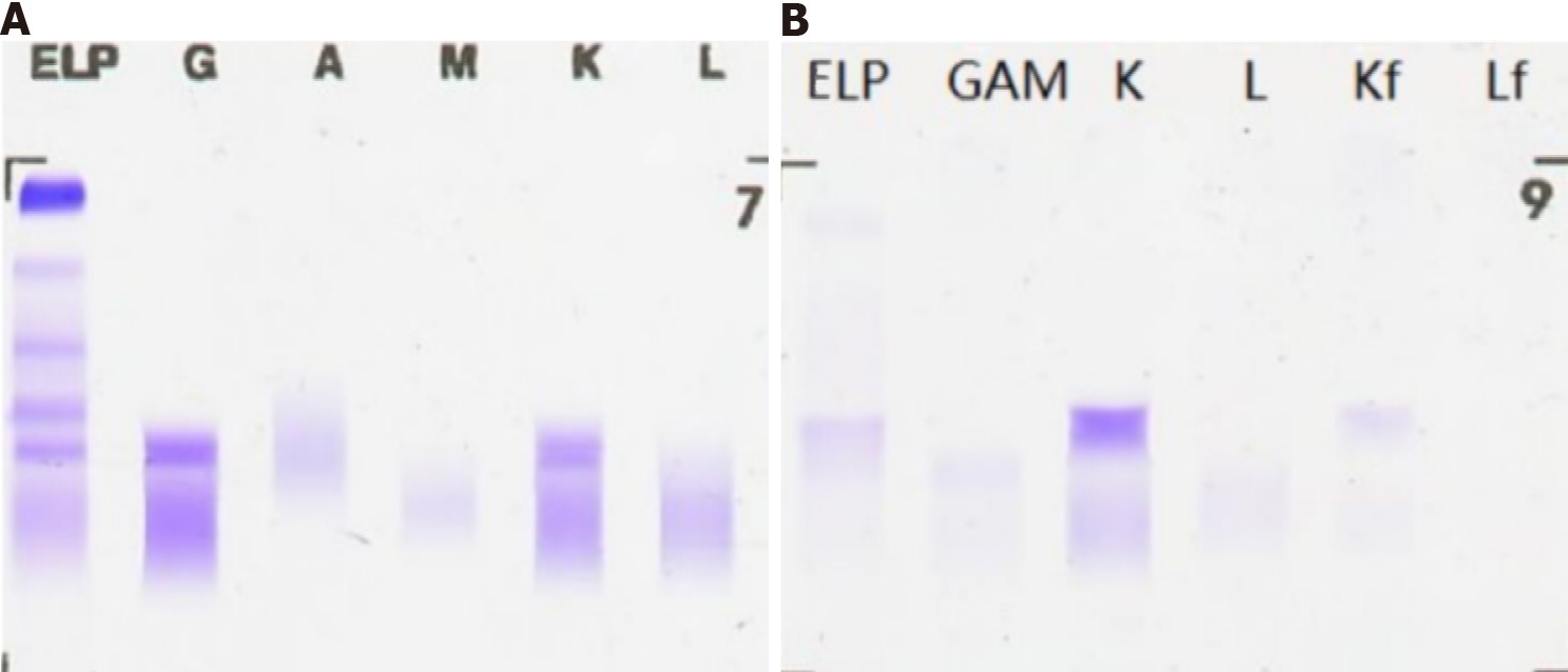
Figure 1 Protein electrophoresis.
A: Serum; B: Urine. ELP: Serum protein electrophoresis; G: Immunoglobulin G; A: Immunoglobulin A; M: Immunoglobulin M; K: Kappa light chain; L: Lambda light chain; Kf: Kappa free light chain; Lf: Lambda free light chain.
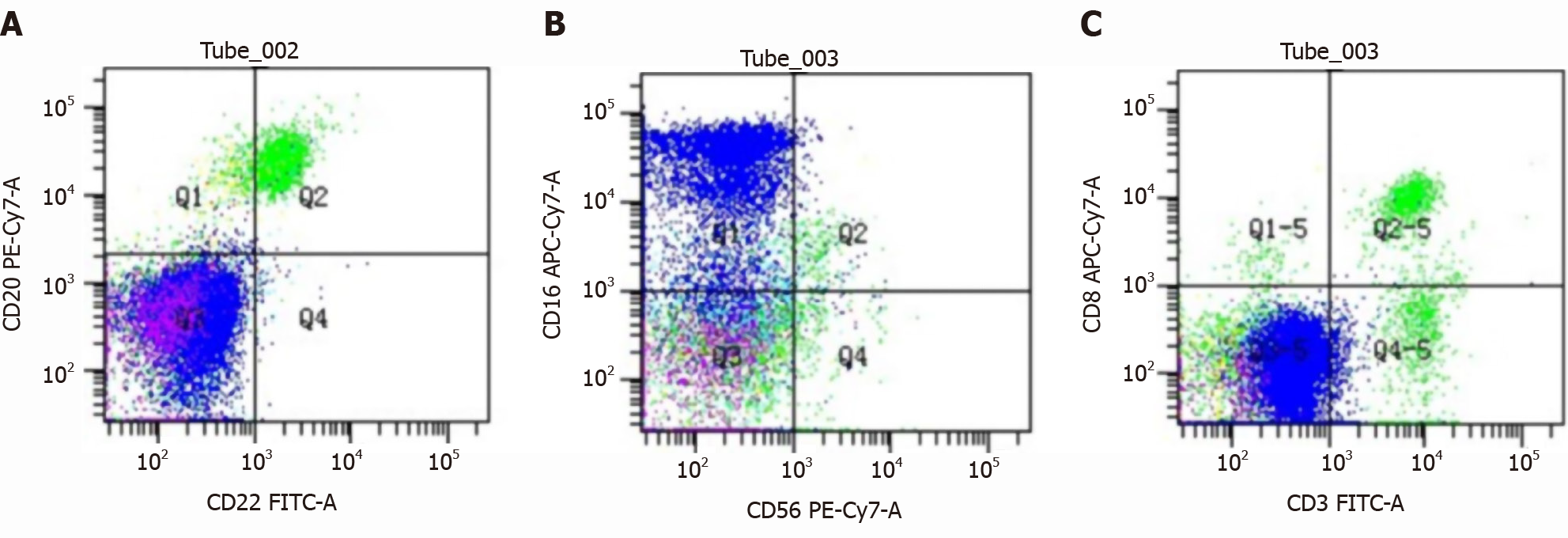
Figure 2 Hematological tumor immunophenotype.
A: CD22 (+) and CD20 (+); B: CD56 (+) and CD16 (+); C: CD3 (+) and CD8 (+).
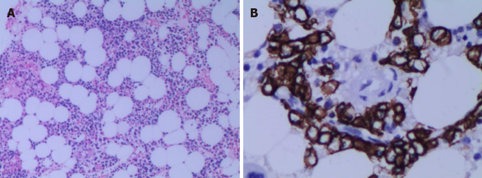
Figure 3 Bone marrow biopsy immunohistochemistry.
Bone marrow biopsy showed active hematopoietic tissue proliferation, scattered granulocytes and thrombocytosis, an increased proportion of erythroblasts, and scattered or small clusters of plasma cells accounted for approximately 5%-10%. It was CD3 (+), CD20 (+), CD56 (-) and CD13 (+). A: Hematoxylin-eosin, 100 ×; B: Immunohistochemistry, 400 ×.
- Citation: Ma Y, Cui S, Yin YJ. Infiltrating ductal breast carcinoma with monoclonal gammopathy of undetermined significance: A case report. World J Clin Cases 2021; 9(15): 3675-3679
- URL: https://www.wjgnet.com/2307-8960/full/v9/i15/3675.htm
- DOI: https://dx.doi.org/10.12998/wjcc.v9.i15.3675


