Copyright
©The Author(s) 2021.
World J Clin Cases. May 16, 2021; 9(14): 3449-3457
Published online May 16, 2021. doi: 10.12998/wjcc.v9.i14.3449
Published online May 16, 2021. doi: 10.12998/wjcc.v9.i14.3449
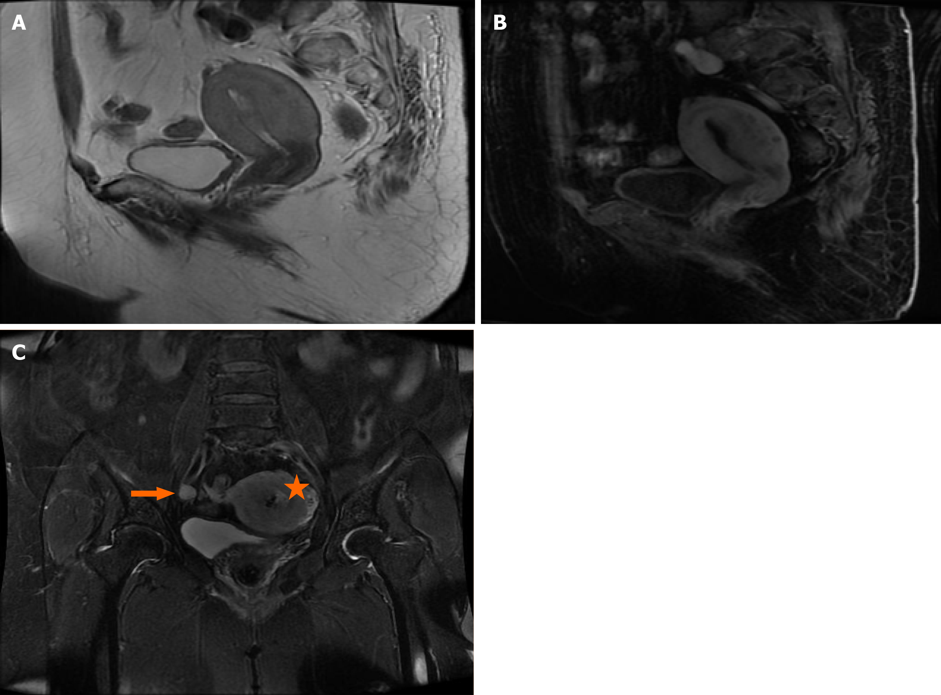
Figure 1 Diagnostic imaging of the tumor.
A: Sagittal T2-weighted magnetic resonance imaging; B: T2-enhanced magnetic resonance imaging of the pelvis revealed an irregular thickened endometrium and a heterogeneously enhanced uterine tumor with diffuse infiltration of the posterior wall of the uterine myometrium; C: Coronal fast spin echo with T2WI fat suppression revealed an enlarged uterus and uterine cavity and local roughness of the serosa (star) and enlargement and partial necrosis of the right internal iliac lymph node (arrows).
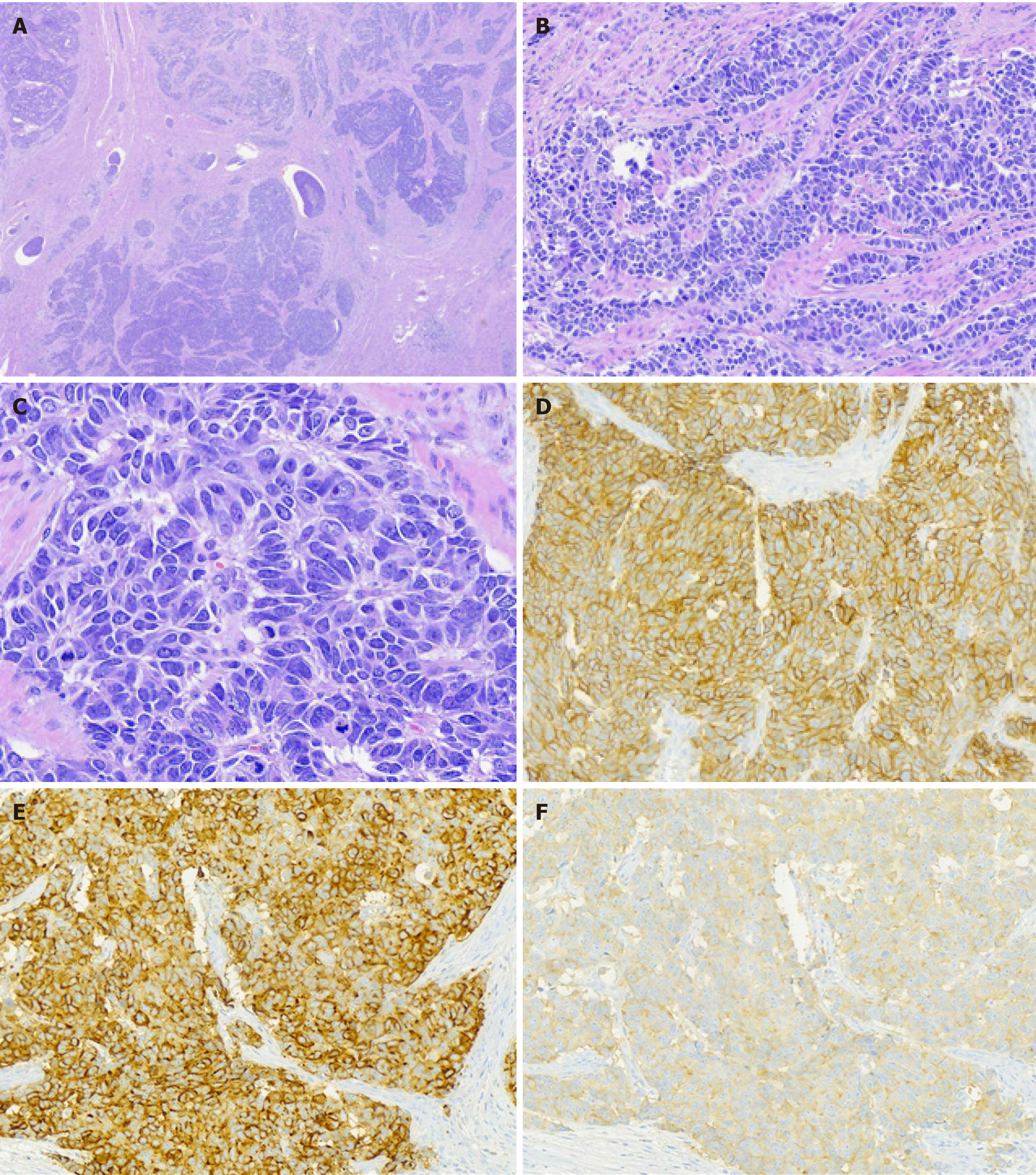
Figure 2 Histology and immunohistochemistry.
A: Microscopically, tumors showed an organoid pattern of growth with tumor embolism (hematoxylin and eosin, × 40); B: Trabeculae-like pattern (hematoxylin and eosin, × 200); C: Rosette-like pattern with “salt and pepper” chromatin (hematoxylin and eosin, × 400); D-F: Immunohistochemistry revealed positivity for CD56 (D, membranous), synaptophysin (E, cytoplasmic), and chromogranin A (F, cytoplasmic).
- Citation: Du R, Jiang F, Wang ZY, Kang YQ, Wang XY, Du Y. Pure large cell neuroendocrine carcinoma originating from the endometrium: A case report. World J Clin Cases 2021; 9(14): 3449-3457
- URL: https://www.wjgnet.com/2307-8960/full/v9/i14/3449.htm
- DOI: https://dx.doi.org/10.12998/wjcc.v9.i14.3449


