Copyright
©The Author(s) 2021.
World J Clin Cases. May 16, 2021; 9(14): 3424-3431
Published online May 16, 2021. doi: 10.12998/wjcc.v9.i14.3424
Published online May 16, 2021. doi: 10.12998/wjcc.v9.i14.3424
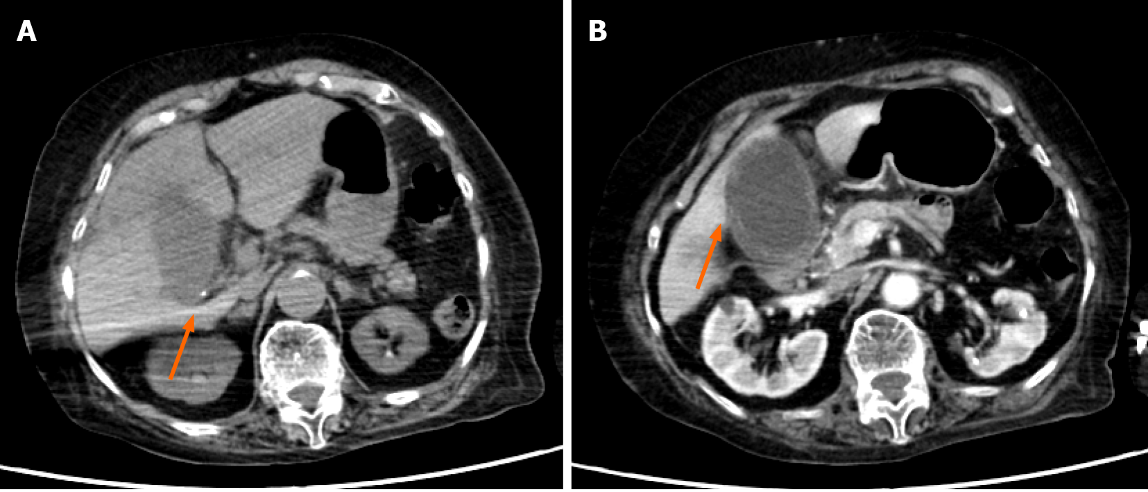
Figure 1 An abdominal contrast-enhanced computed tomography (Case 1).
A: Small calculus in the gallbladder neck (arrow); B: Significant swelling of the gallbladder and asymmetrical wall thickening. The gallbladder wall indicated parts with either poor or no contrast enhancement (arrow), with a partial failure of continuity of the mucosa.
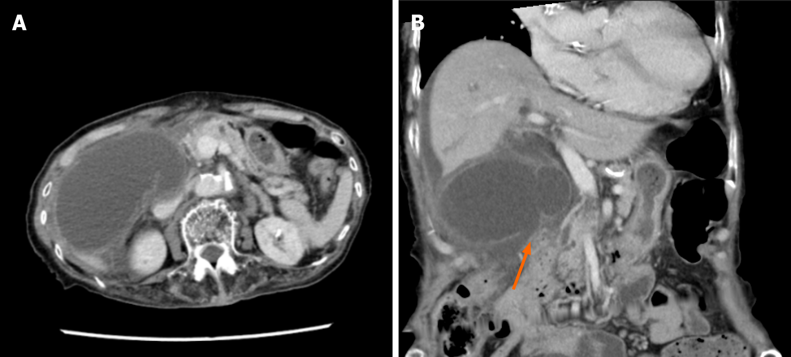
Figure 2 An abdominal contrast-enhanced computed tomography (Case 2).
A: Significant swelling of the gallbladder. Ascites around the liver; B: There was asymmetrical gallbladder wall thickening and the continuity of part of the wall was ambiguous (arrow).
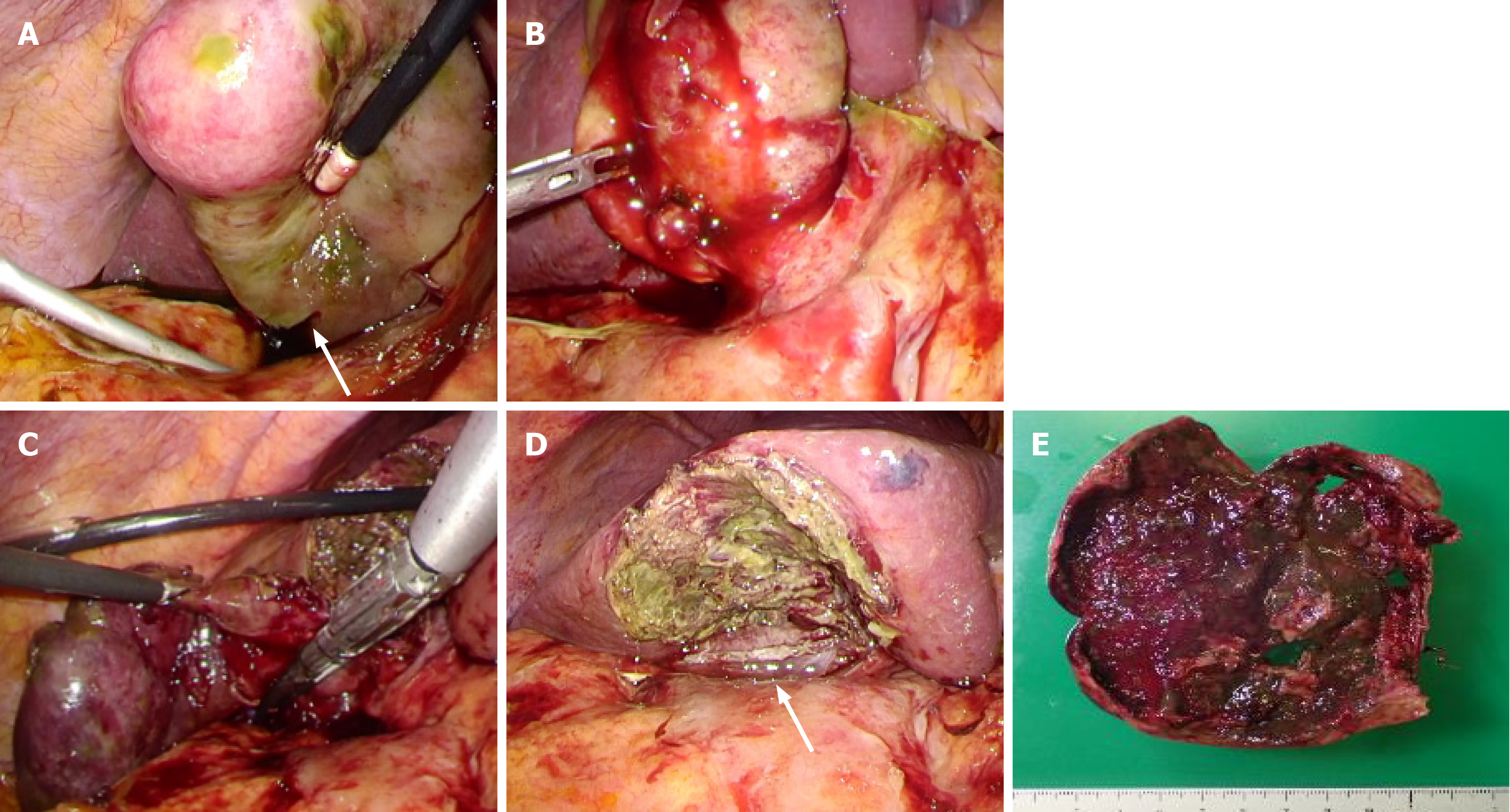
Figure 3 Surgical observations and surgical techniques (Case 1).
A: Gallbladder necroses and a part thereof is perforated (arrow), with leaking of bile; B: Gallbladder neck had substantial inflammation; C: Upon exfoliation from the base to the neck of the gallbladder, we treated the neck using an autosuture; D: After subtotal resection of the gallbladder, the arrow is the amputation stump of the gallbladder neck; E: Resected specimen.
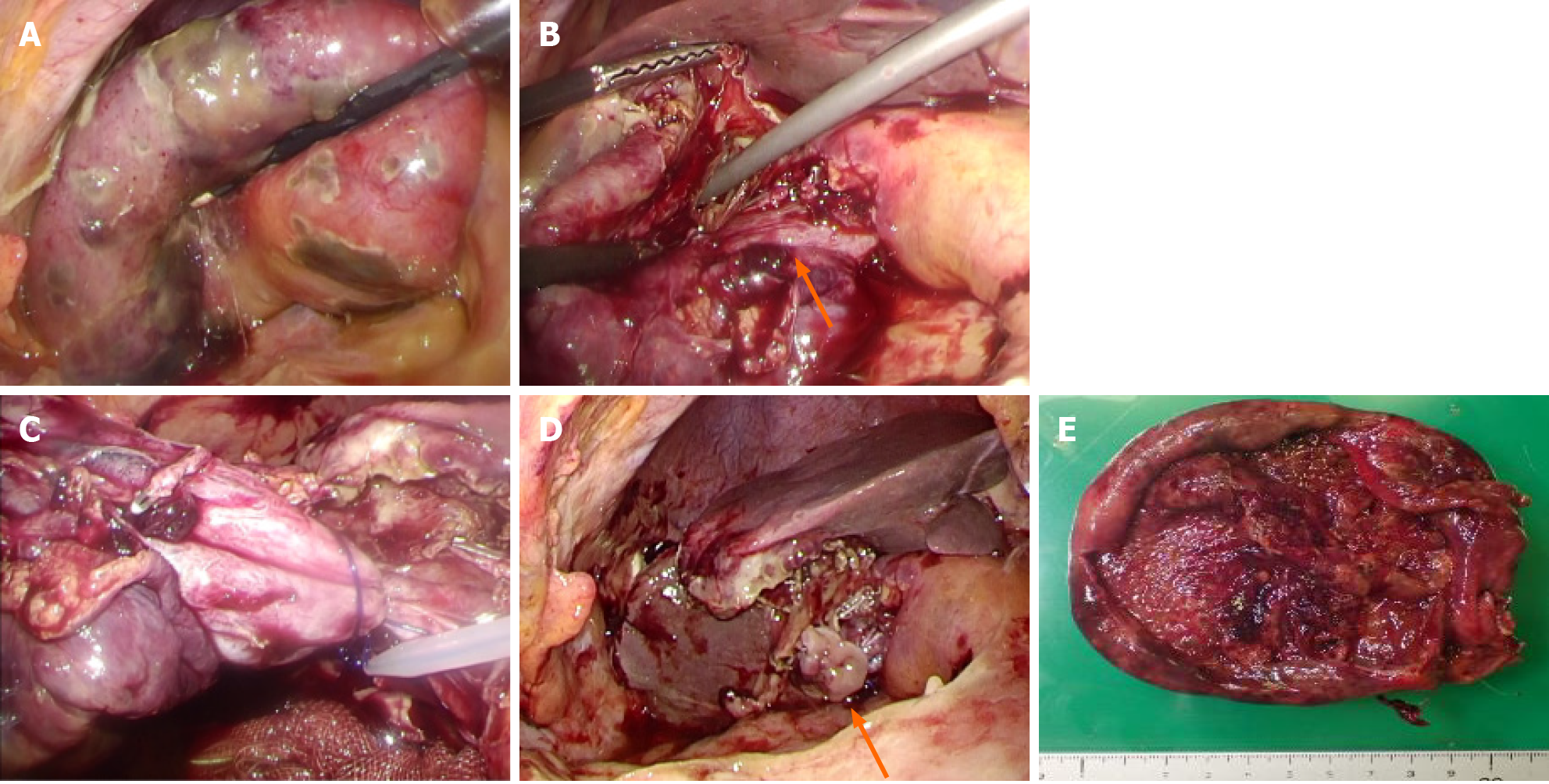
Figure 4 Surgical observations and surgical techniques (Case 2).
A: The gallbladder had significant swelling and diffuse necrosis; B: The arrow is a cystic duct; C: For the cystic duct and gallbladder neck, we performed ligature/treatment using a looped absorbable monofilament suture; D: After subtotal resection of the gallbladder, the arrow is the amputation stump of the gallbladder neck; E: Resected specimen.
- Citation: Inoue H, Ochiai T, Kubo H, Yamamoto Y, Morimura R, Ikoma H, Otsuji E. Laparoscopic cholecystectomy for gangrenous cholecystitis in around nineties: Two case reports. World J Clin Cases 2021; 9(14): 3424-3431
- URL: https://www.wjgnet.com/2307-8960/full/v9/i14/3424.htm
- DOI: https://dx.doi.org/10.12998/wjcc.v9.i14.3424


