Copyright
©The Author(s) 2021.
World J Clin Cases. May 16, 2021; 9(14): 3394-3402
Published online May 16, 2021. doi: 10.12998/wjcc.v9.i14.3394
Published online May 16, 2021. doi: 10.12998/wjcc.v9.i14.3394
Figure 1 Preoperative X-ray, computed tomography, and magnetic resonance imaging images.
A: Plain X-ray of the cervical spine showing destructive lesions (orange arrow) in the appendage area of the C4-5 vertebras; B: Plain computed tomography (CT) of the cervical spine showing bone destruction (orange arrow) and a soft tissue mass in the appendage area of the C4-5 vertebras; C: CT three-dimensional imaging of the cervical spine showing the outline of bone destruction (orange arrow) in the appendage area of C4-5; D: Magnetic resonance imaging showing bone destruction and soft tissue formation (orange arrow) in the C4-C5 accessory region, suggesting the possibility of an invasive, benign bone tumor (e.g., osteoblastoma), and significant compression of the spinal cord and stenosis at the corresponding plane.
Figure 2 Intraoperative images.
A: Operative view of the lesion between the C3 and C6 spinous process lamina; B: View of the surgeon after lesion removal and spinal canal and nerve root canal decompression; C: Nodular fragment of tissue, measuring 7.2 cm × 6.5 cm × 5.4 cm after resection.
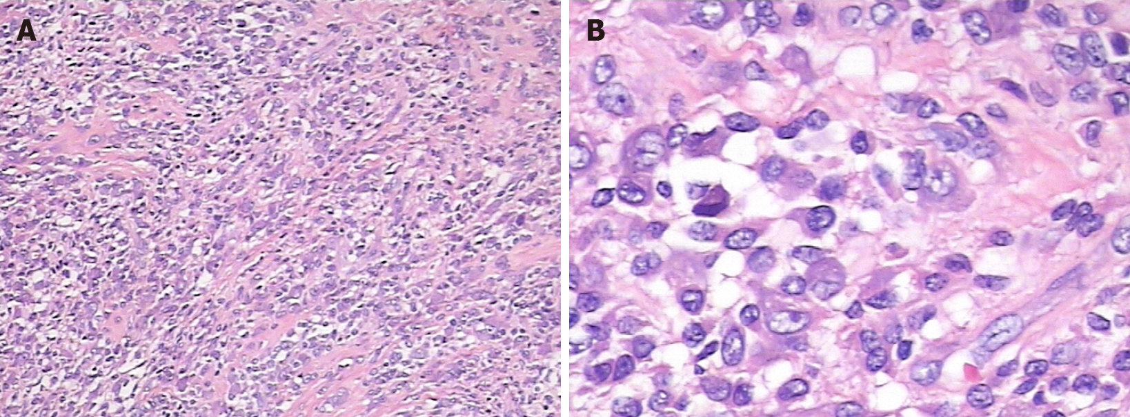
Figure 3 Pathological images of the mass.
The histopathological analysis mainly showed proliferating monocytes and osteoclastic multinucleated giant cells. A: Image at 40 × magnification; B: Image at 200 × magnification.
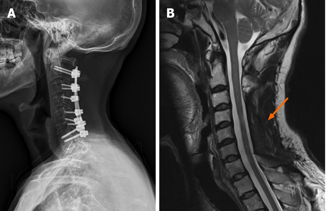
Figure 4 X-ray and magnetic resonance imaging review images at 1 year postoperatively.
A: X-ray showing good fixation with no loosening of the internal fixation; B: Magnetic resonance imaging showing no signs of recurrence (orange arrow).
- Citation: Zhu JH, Li M, Liang Y, Wu JH. Tenosynovial giant cell tumor involving the cervical spine: A case report . World J Clin Cases 2021; 9(14): 3394-3402
- URL: https://www.wjgnet.com/2307-8960/full/v9/i14/3394.htm
- DOI: https://dx.doi.org/10.12998/wjcc.v9.i14.3394


