Copyright
©The Author(s) 2021.
World J Clin Cases. May 16, 2021; 9(14): 3379-3384
Published online May 16, 2021. doi: 10.12998/wjcc.v9.i14.3379
Published online May 16, 2021. doi: 10.12998/wjcc.v9.i14.3379
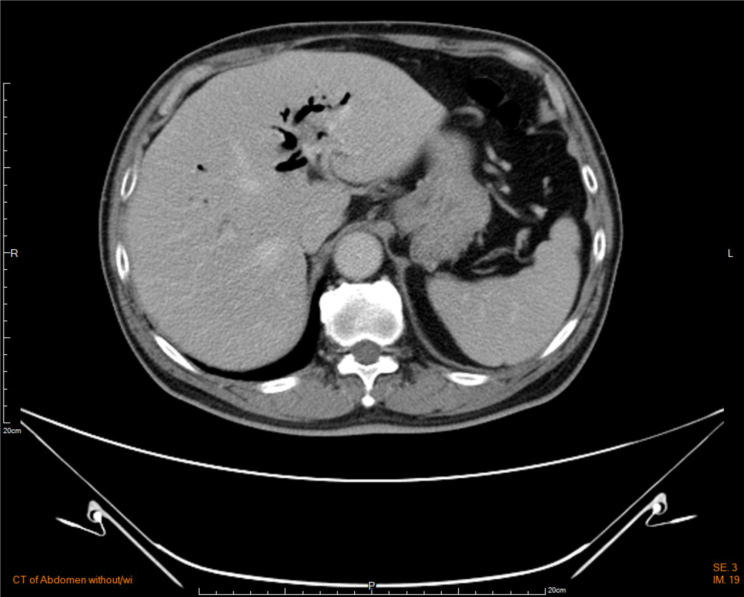
Figure 1 Computed tomography scan of the abdomen demonstrated gas retention in the intrahepatic ducts, suggesting pneumobilia.
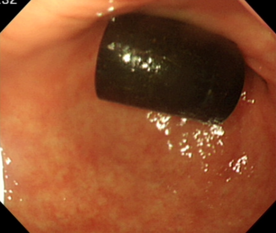
Figure 2 There was a capsule-like foreign body approximately 1 cm × 2 cm in size at the gastric antrum and peri-pyloric region.
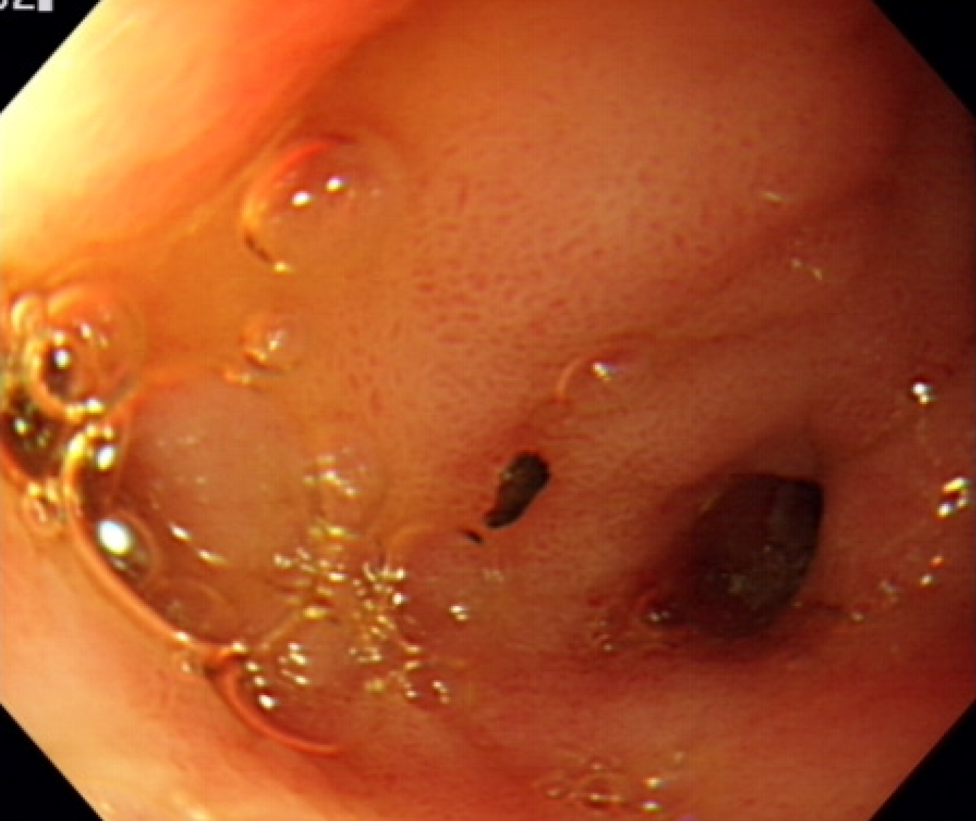
Figure 3 Orifice present over the pyloric ring in stomach noted after removal of a foreign body.
An ectopic ampulla of Vater was suspected.
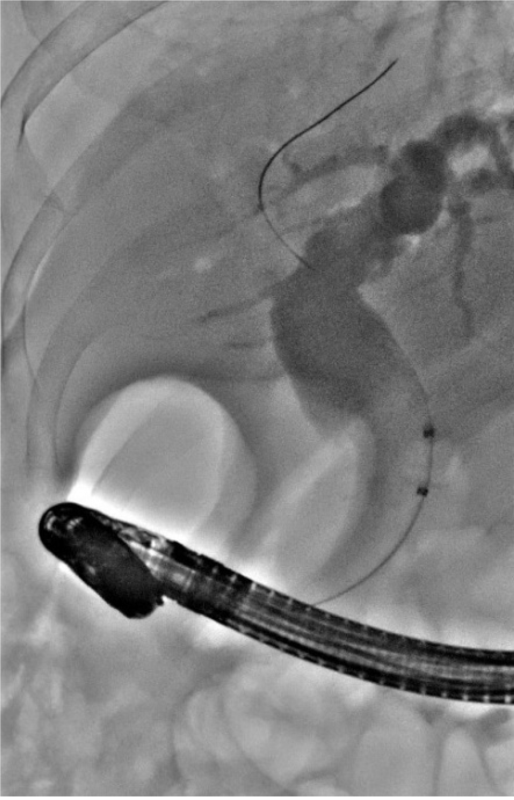
Figure 4 Cholangiography.
The wire-guided catheter was inserted through the endoscope into the ectopic orifice at the pyloric ring.
Figure 5 T1-weighted magnetic resonance images with gadolinium-based contrast media.
A: Common bile duct (arrow) was dilated; B and C: Common bile duct (arrow) narrowed and drained into the pylorus.
- Citation: Lee HL, Fu CK. Acute cholangitis detected ectopic ampulla of Vater in the antrum incidentally: A case report. World J Clin Cases 2021; 9(14): 3379-3384
- URL: https://www.wjgnet.com/2307-8960/full/v9/i14/3379.htm
- DOI: https://dx.doi.org/10.12998/wjcc.v9.i14.3379


