Copyright
©The Author(s) 2021.
World J Clin Cases. May 16, 2021; 9(14): 3372-3378
Published online May 16, 2021. doi: 10.12998/wjcc.v9.i14.3372
Published online May 16, 2021. doi: 10.12998/wjcc.v9.i14.3372
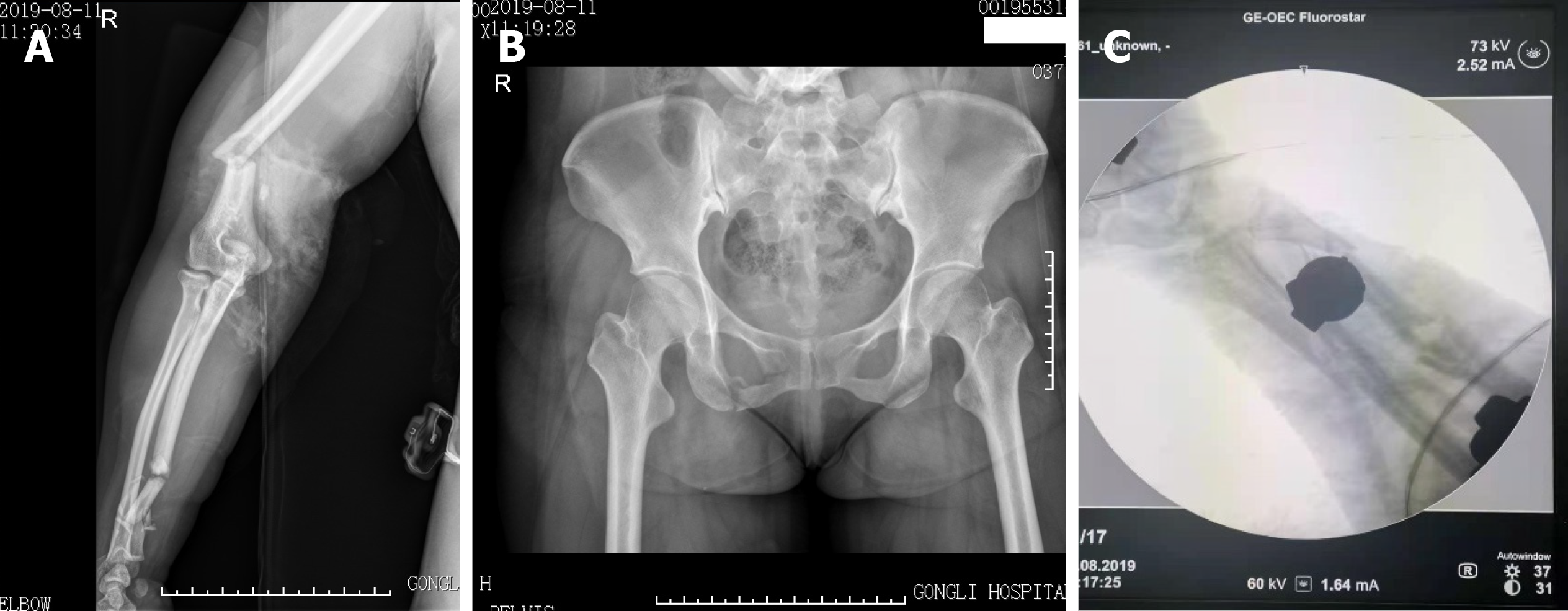
Figure 1 X-ray images of the patient’s pelvic fracture.
A: X-ray image showed the fracture of the distal right humerus, right olecranon and multiple segment; B: X-ray image showed the fracture of the pelvis; C: X-ray from the C-arm fluoroscopic machine showed comminuted fractures of the ulna and radius.
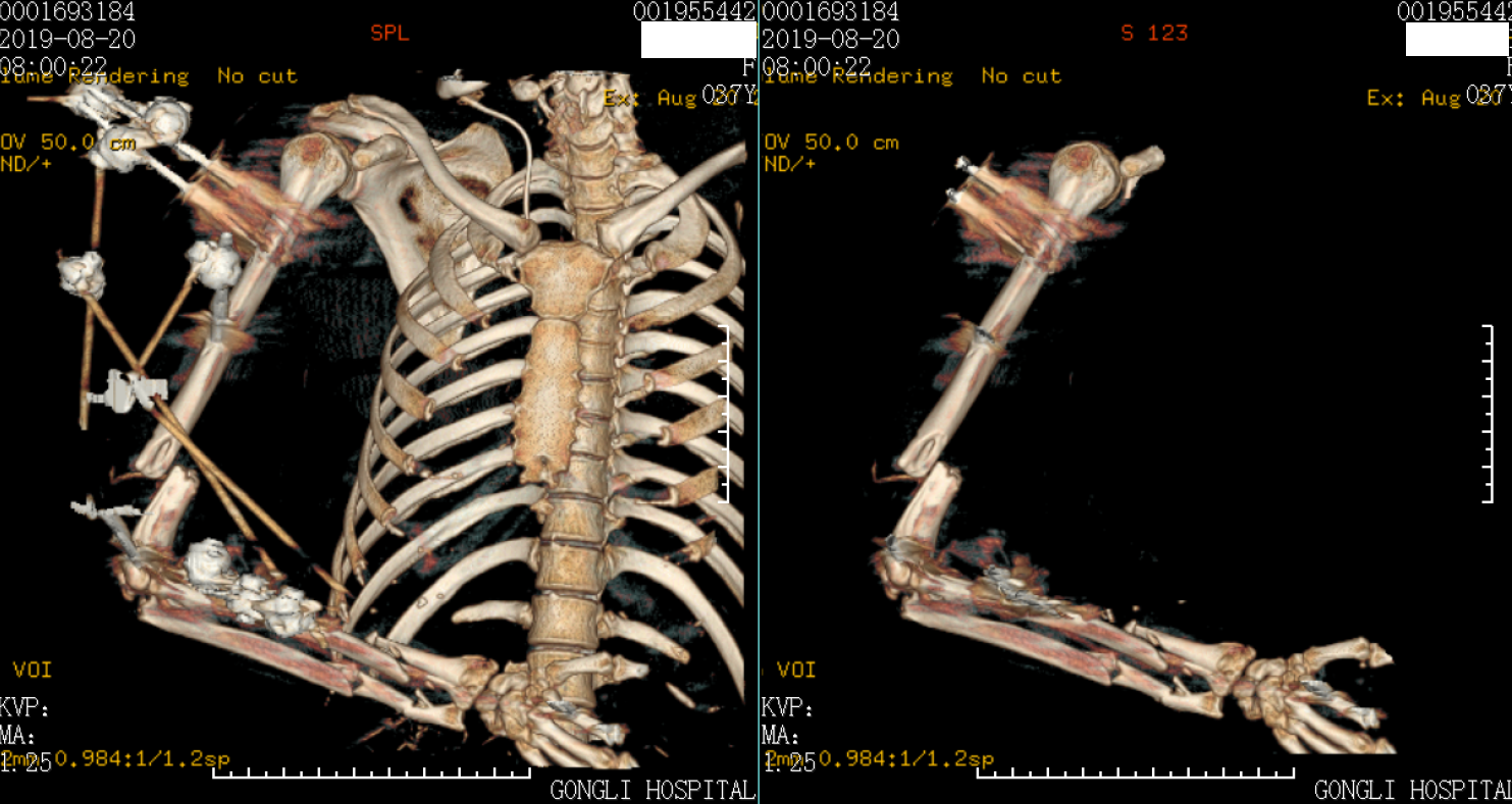
Figure 2 Three-dimensional computed tomography scans of the patient with external fixators.
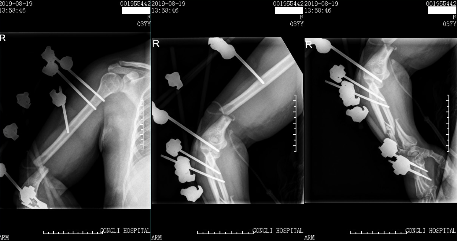
Figure 3 X-ray images of the patient with external fixators.
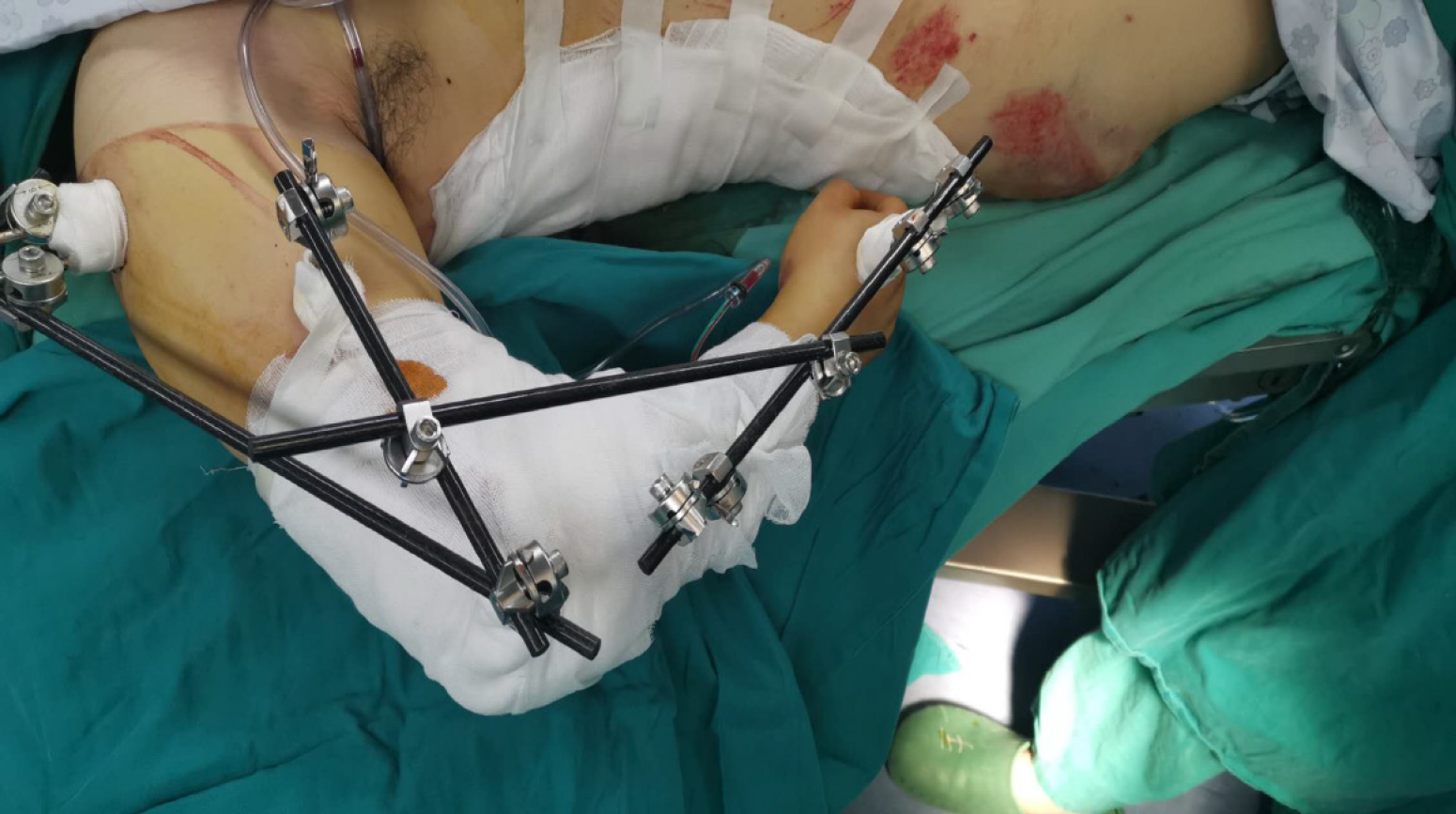
Figure 4 Image of the patient after the first emergency operation.
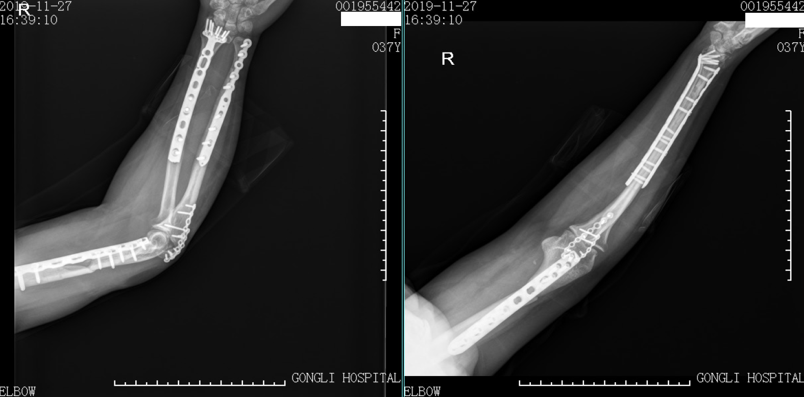
Figure 5 X-ray images of the patient at the 7 mo follow-up.
Figure 6 Movements of the patient at the 7 mo follow-up.
- Citation: Huang GH, Tang JA, Yang TY, Liu Y. Floating elbow combining ipsilateral distal multiple segmental forearm fractures: A case report. World J Clin Cases 2021; 9(14): 3372-3378
- URL: https://www.wjgnet.com/2307-8960/full/v9/i14/3372.htm
- DOI: https://dx.doi.org/10.12998/wjcc.v9.i14.3372


