Copyright
©The Author(s) 2021.
World J Clin Cases. May 16, 2021; 9(14): 3356-3364
Published online May 16, 2021. doi: 10.12998/wjcc.v9.i14.3356
Published online May 16, 2021. doi: 10.12998/wjcc.v9.i14.3356
Figure 1 Initial axial non-contrast computed tomography of the head.
A: Extravasated blood predominantly in the perimesencephalic subarachnoid space within the extension into bilateral ambient cisterns; B: Extravasated blood predominantly in the quadrigeminal cistern and chiasmatic cisterns; C: Extravasated blood predominantly in the quadrigeminal cistern and chiasmatic cisterns; D: The third ventricle was slightly ectatic.
Figure 2 Cerebral angiography on the day of admission.
A: Right vertebral artery angiography; B: Lateral angiography of the right vertebral artery; C: Anterior angiography of the left vertebral artery; D: Lateral angiography of the left vertebral artery; E: Right internal carotid artery angiography; F: Lateral angiography of the right internal carotid artery; G: Angiography of the left internal carotid artery; H: Lateral angiography of the left internal carotid artery; I: Right external carotid artery angiography anteroposterior; J: Lateral angiography of the right external carotid artery; K: External left carotid artery angiography anteroposterior; L: Lateral angiography of the left external carotid artery.
Figure 3 After lumbar puncture, blood was dissolved.
Four days after the initial bleeding, non-contrast computed tomography (CT) showed the typical pattern of perimesencephalic spontaneous subarachnoid hemorrhage (SAH). A: After lumbar puncture, blood was dissolved; B: CT shows blood was dissolved; C: The typical pattern of perimesencephalic SAH; D: The typical pattern of perimesencephalic SAH.
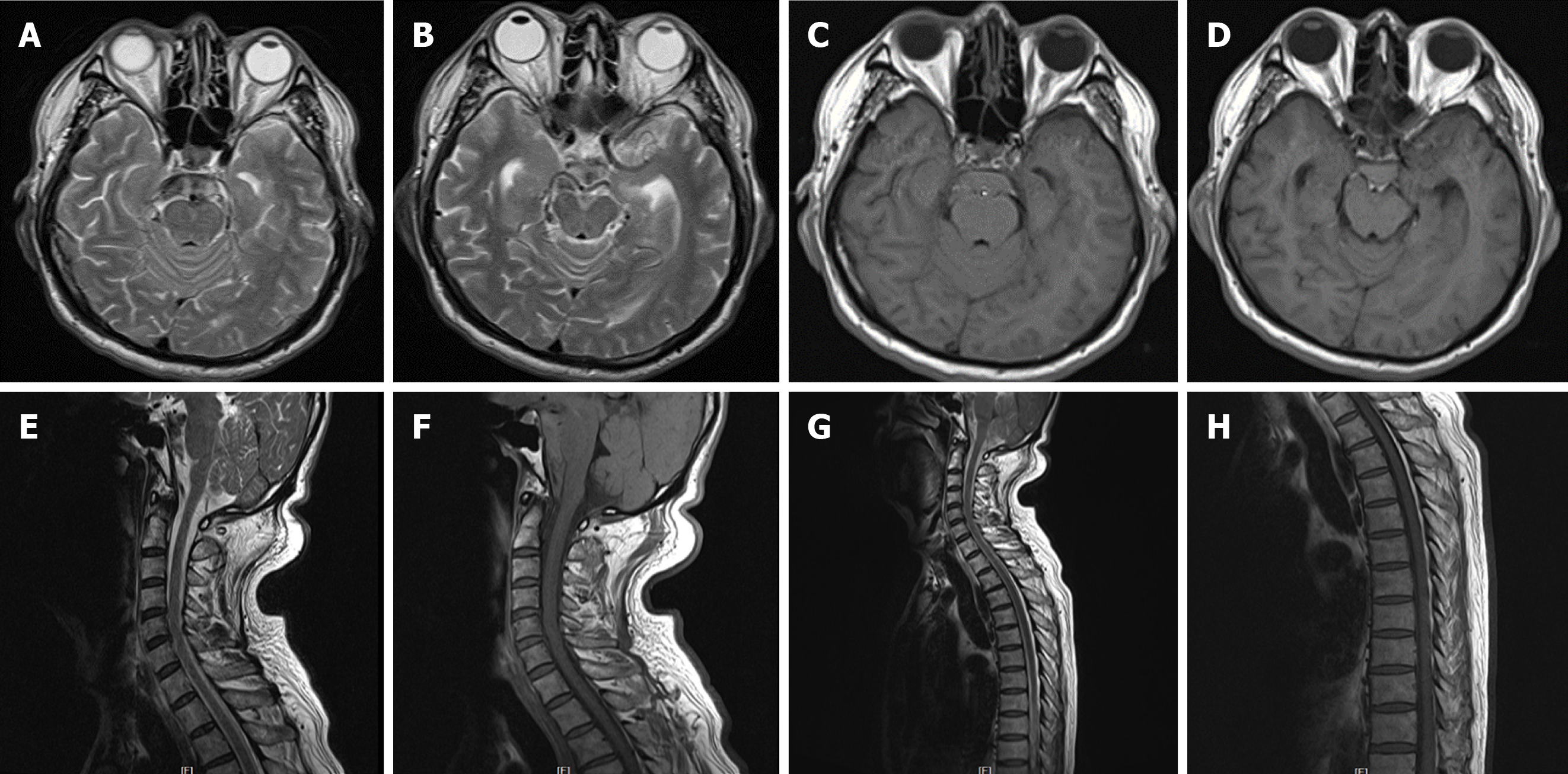
Figure 4 Magnetic resonance imaging of the brain, cervical spinal cord, and upper thoracic spinal cord performed the following day after second hemorrhage.
A and B: Axial T2 weighted imaging (T2WI) of the brain showed hyperintense signal; C and D: T1 weighted imaging (T1WI) exhibited equal intense signals in the perimesencephalic cisterns; E-H: Underlying structural abnormalities were not revealed. Sagittal T2WI and T1WI of the cervical and upper thoracic spinal cord were unremarkable.
Figure 5 Six-vessel cerebral catheter angiograms of bilateral thyrocervical and costocervical trunks ruling them out as sources of spontaneous subarachnoid hemorrhage.
A and B: Bilateral vertebral artery injection [arterial phase; anteroposterior (AP) projection]; C and D: Bilateral internal carotid artery injection (arterial phase; AP projection); E and F: Right internal carotid artery injection (venous phase; AP and lateral projections); G and H: Left internal carotid artery injection (venous phase; AP and lateral projections); I and J: Right thyrocervical trunk injection (arterial phase; AP and lateral projections); K and L: Left thyrocervical trunk injection (arterial phase; AP and lateral projections). Internal carotid artery angiography illustrated the drainage of basal vein of Rosenthal (BVR), which does not drain into the vein of Galen due to discontinuous BVR (E-H).
- Citation: Li J, Fang X, Yu FC, Du B. Recurrent perimesencephalic nonaneurysmal subarachnoid hemorrhage within a short period of time: A case report. World J Clin Cases 2021; 9(14): 3356-3364
- URL: https://www.wjgnet.com/2307-8960/full/v9/i14/3356.htm
- DOI: https://dx.doi.org/10.12998/wjcc.v9.i14.3356


