Copyright
©The Author(s) 2021.
World J Clin Cases. May 16, 2021; 9(14): 3334-3341
Published online May 16, 2021. doi: 10.12998/wjcc.v9.i14.3334
Published online May 16, 2021. doi: 10.12998/wjcc.v9.i14.3334
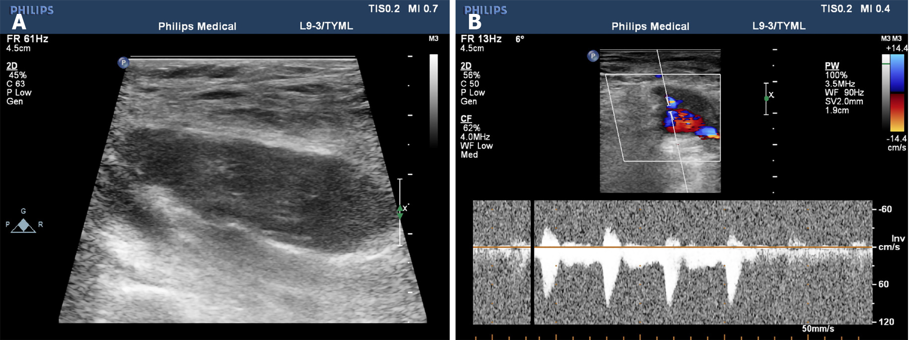
Figure 1 Imaging examinations.
A: Two-dimensional ultrasound images show mixed echoes in the muscularis of the right thigh with unclear boundaries; B: Power Doppler activity revealed significantly increased flow within the tumor.
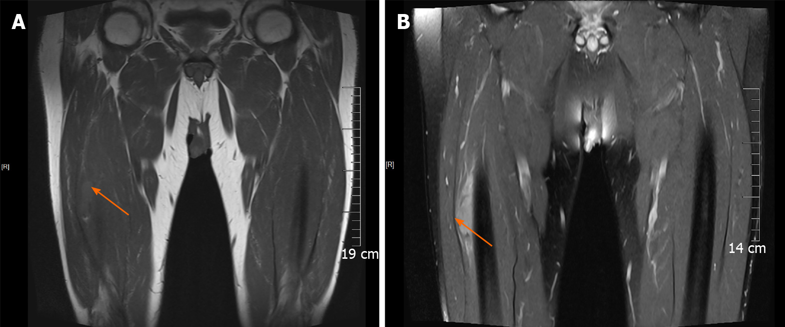
Figure 2 Magnetic resonance imaging.
A: Coronal scan of the arrow points iso-signal of the tumor intensity on T1-weighted images; B: The arrow indicates high-signal intensity on T2-weighted images compared with the surrounding muscle.
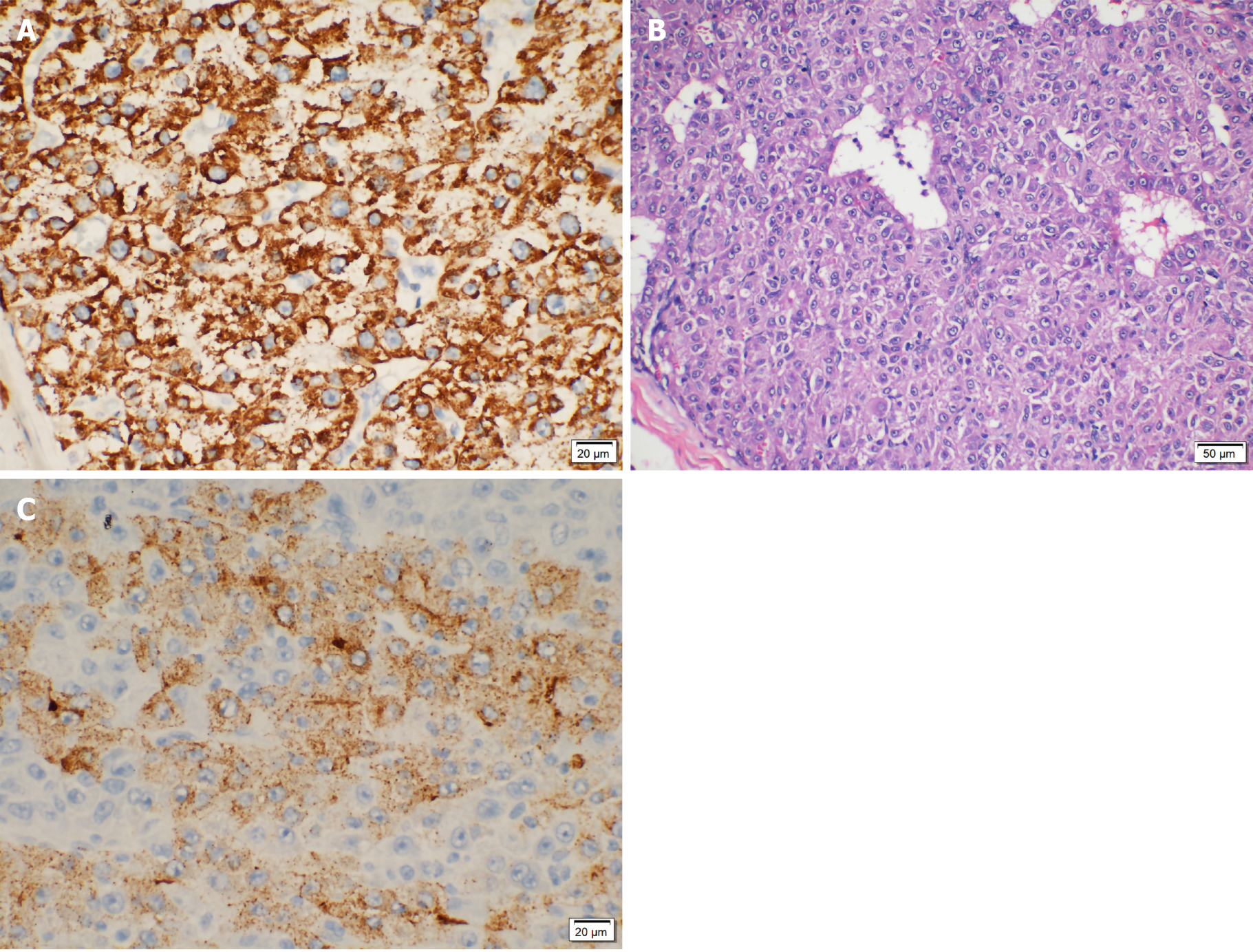
Figure 3 Pathological analysis.
A: Immunohistochemical staining examination of metastatic hepatic carcinoma hepatocyte paraffine 1 (Stain, 400 ×); B: Pathological examination of the tumor in the right thigh (Hematoxylin stain, 200 ×); C: Immunohistochemical staining examination of the tumor in the right thigh of metastatic hepatic carcinoma glypican-3 (Stain, 400 ×).
- Citation: Song Q, Sun XF, Wu XL, Dong Y, Wang L. Skeletal muscle metastases of hepatocellular carcinoma: A case report and literature review . World J Clin Cases 2021; 9(14): 3334-3341
- URL: https://www.wjgnet.com/2307-8960/full/v9/i14/3334.htm
- DOI: https://dx.doi.org/10.12998/wjcc.v9.i14.3334


