Copyright
©The Author(s) 2021.
World J Clin Cases. May 16, 2021; 9(14): 3327-3333
Published online May 16, 2021. doi: 10.12998/wjcc.v9.i14.3327
Published online May 16, 2021. doi: 10.12998/wjcc.v9.i14.3327
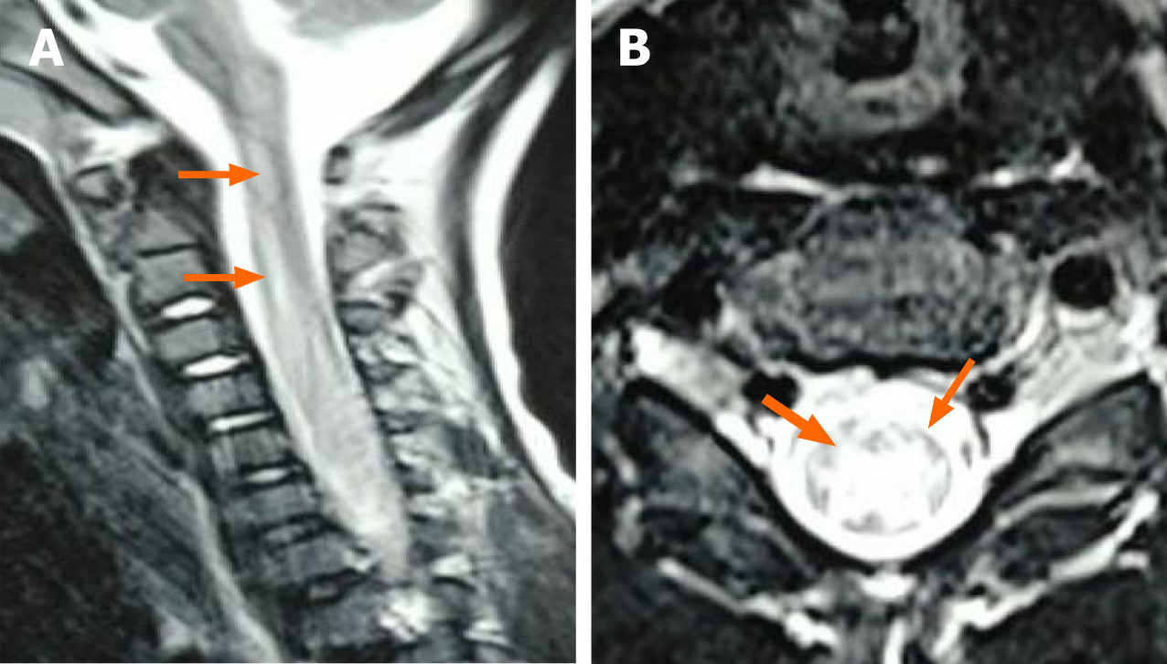
Figure 1 Magnetic resonance imaging of the cervical spinal cord in case 1.
A: T2 high signal shadow in the anterior horn at levels C2-C6 of the spinal cord; B: Signals are more obvious on the left side, considering the presence of inflammation.
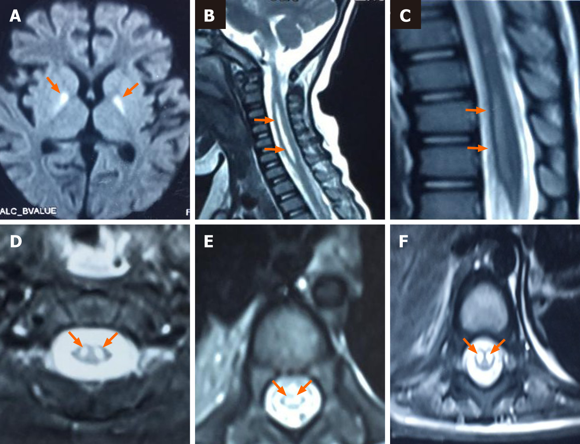
Figure 2 Magnetic resonance imaging of the brain and spinal cord in case 2.
A: Abnormal signals in the bilateral basal ganglia; B-F: T2 high signal shadow in the anterior horn at levels C1-T1 and T8-L1 of the spinal cord.
- Citation: Zhang Y, Wang SY, Guo DZ, Pan SY, Lv Y. Acute flaccid paralysis and neurogenic respiratory failure associated with enterovirus D68 infection in children: Report of two cases. World J Clin Cases 2021; 9(14): 3327-3333
- URL: https://www.wjgnet.com/2307-8960/full/v9/i14/3327.htm
- DOI: https://dx.doi.org/10.12998/wjcc.v9.i14.3327


