Copyright
©The Author(s) 2021.
World J Clin Cases. May 16, 2021; 9(14): 3320-3326
Published online May 16, 2021. doi: 10.12998/wjcc.v9.i14.3320
Published online May 16, 2021. doi: 10.12998/wjcc.v9.i14.3320
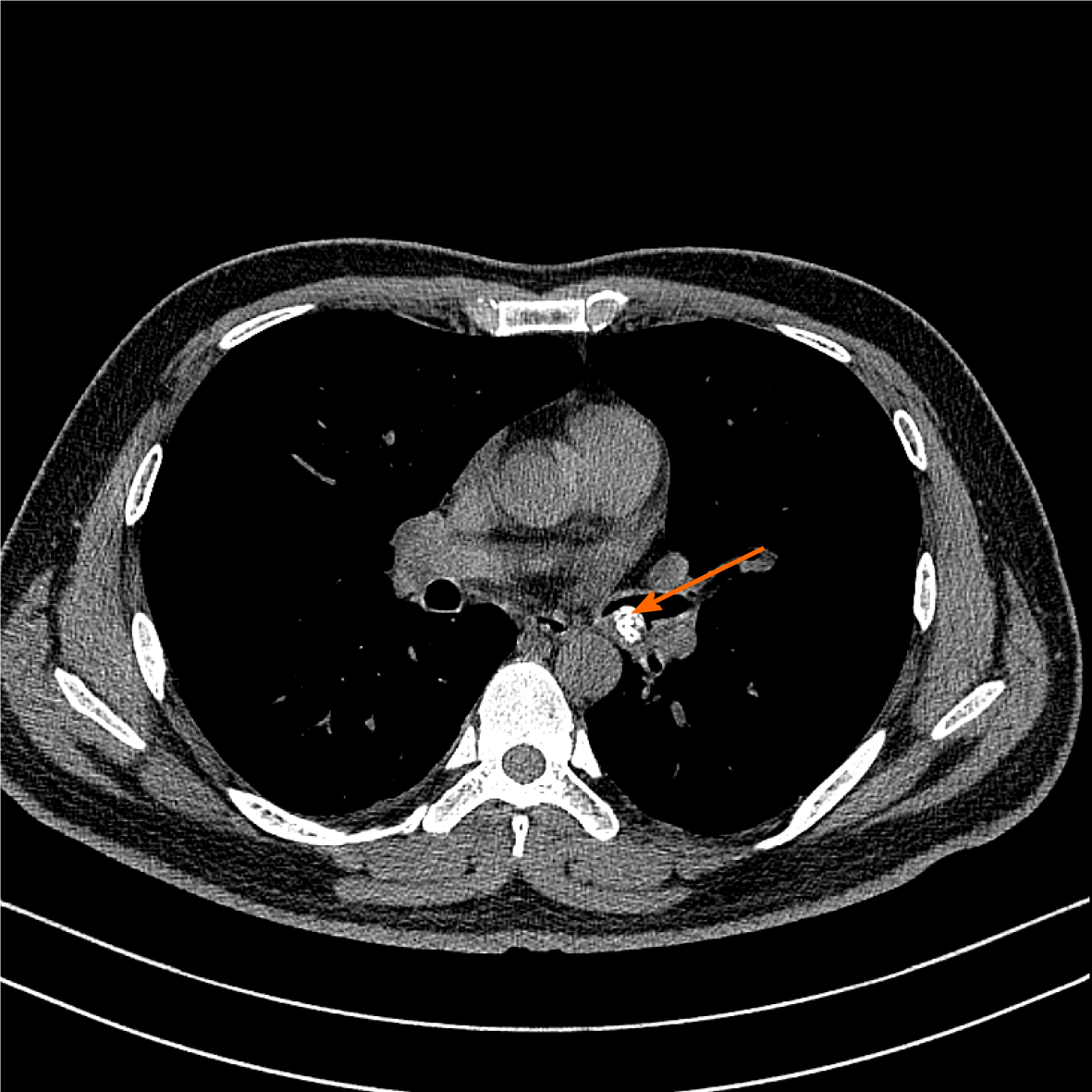
Figure 1 Chest computed tomography.
A 1.20 cm × 0.88 cm calcified nodular lesion on the compressed posterior wall of the lower left main bronchus (orange arrow).
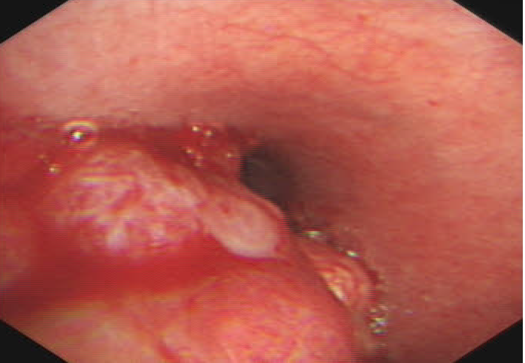
Figure 2 Bronchoscopic view of basal segment of the lower left lobe.
A yellow–white mass obstructed the entrance to the basal segment of the lower left lobe. Lateral to the entrance of the basal segment of the lower left lobe, neoplasms with multiple nodular ridges and superficial hyperemia were observed.
Figure 3 Bronchial glomus tumor histopathology.
A: Tumor cells were uniformly round with smooth nuclear contours, fine chromatin and a modest amount of pink cytoplasm. They were arranged in sheet-like patterns between small blood vessels (hematoxylin–eosin staining, 200×); B: Tumor cells were positive for smooth muscle actin (immunohistochemical staining, 400×); C: Tumor cells were positive for actin(immunohistochemical staining, 400×).
- Citation: Zhang Y, Zhang QP, Ji YQ, Xu J. Bronchial glomus tumor with calcification: A case report. World J Clin Cases 2021; 9(14): 3320-3326
- URL: https://www.wjgnet.com/2307-8960/full/v9/i14/3320.htm
- DOI: https://dx.doi.org/10.12998/wjcc.v9.i14.3320


