Copyright
©The Author(s) 2021.
World J Clin Cases. May 6, 2021; 9(13): 3194-3199
Published online May 6, 2021. doi: 10.12998/wjcc.v9.i13.3194
Published online May 6, 2021. doi: 10.12998/wjcc.v9.i13.3194
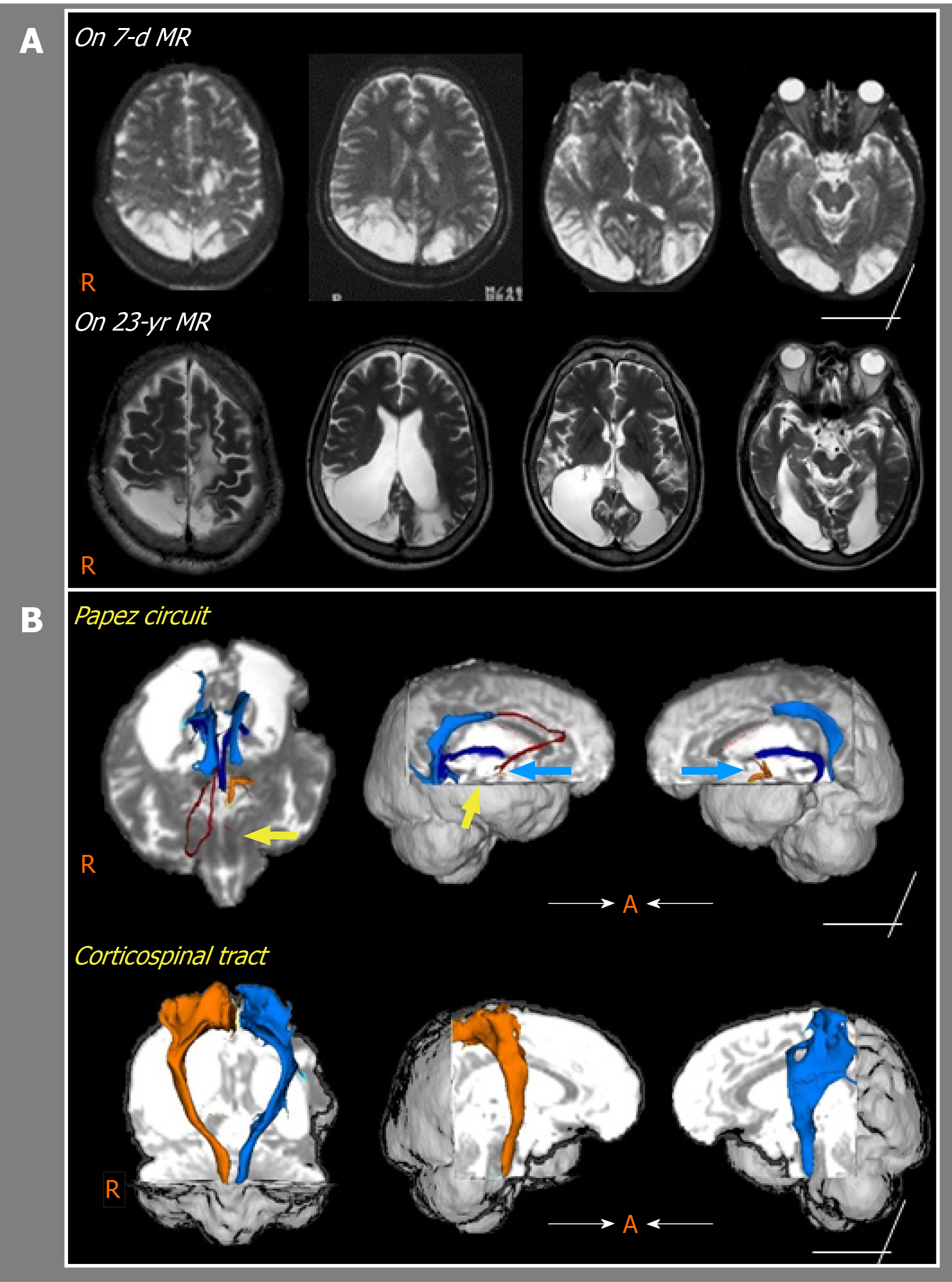
Figure 1 Serial magnetic resonance imaging and diffusion tensor tractography of a chronic patient with cognitive impairment after scrub typhus encephalitis.
A: Magnetic resonance imaging shows multiple lesions in the parietal, temporal, and occipital lobes of both hemispheres. Compared with the findings of the 7-d magnetic resonance imaging, the 23-yr magnetic resonance imaging findings indicates expanded lesions of encephalomalacic changes with marked dilation of both ventricles; B: Diffusion tensor tractography of the Papez circuit and corticospinal tract reveals discontinued of the left thalamocortical and right mammillothalamic tracts in the Papez circuit (yellow arrows). In addition, the anterior part of the fornix is not observed in both hemispheres (blue arrows). The corticospinal tracts of both hemispheres appear relatively intact. MR: Magnetic resonance.
- Citation: Kwon HG, Yang JH, Kwon JH, Yang D. Association between scrub typhus encephalitis and diffusion tensor tractography detection of Papez circuit injury: A case report. World J Clin Cases 2021; 9(13): 3194-3199
- URL: https://www.wjgnet.com/2307-8960/full/v9/i13/3194.htm
- DOI: https://dx.doi.org/10.12998/wjcc.v9.i13.3194


