Copyright
©The Author(s) 2021.
World J Clin Cases. Apr 26, 2021; 9(12): 2874-2883
Published online Apr 26, 2021. doi: 10.12998/wjcc.v9.i12.2874
Published online Apr 26, 2021. doi: 10.12998/wjcc.v9.i12.2874
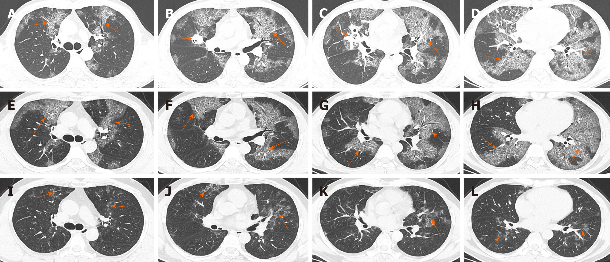
Figure 1 Lung computed tomography findings.
A-D: In August 2018, diffuse interstitial changes were observed within the lung, with local consolidation in the right middle lobe (arrow); E-H: In April 2019, significant absorption of the consolidated area in the middle lobe of the right lung was observed, while diffuse interstitial changes were largely unchanged (arrow); I-L: In December 2019, significant absorption of the diffuse bilateral interstitial changes was observed (arrow).
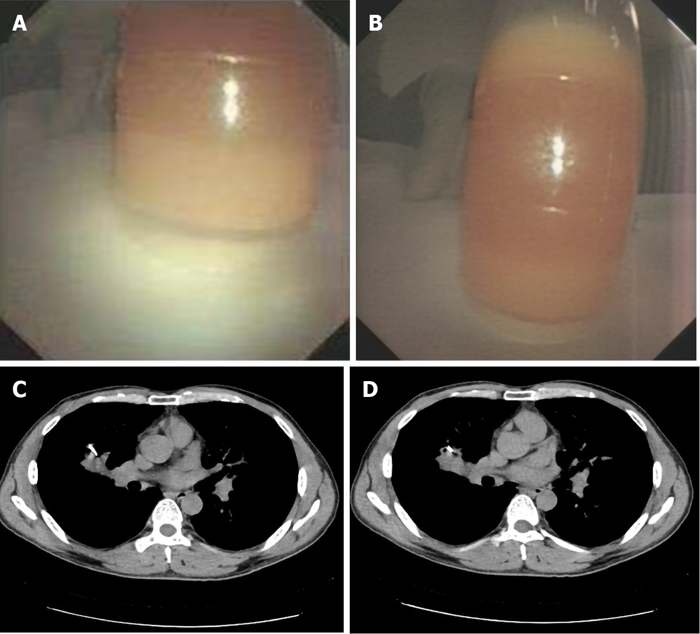
Figure 2 Bronchoalveolar lavage fluid and computed tomography-guided lung biopsy.
A and B: The bronchoalveolar lavage fluid appeared milky white and turbid; C and D: Computed tomography revealed the location of the consolidation in the middle lobe of the right lung.
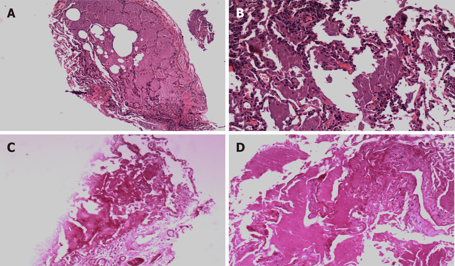
Figure 3 Transbronchial lung biopsy pathology.
A and B: Hematoxylin-eosin-stained lung biopsied tissues (A: 40 ×; B: 100 ×); C and D: Periodic acid-Schiff-stained lung biopsied tissues (C: 40 ×; D: 100 ×).
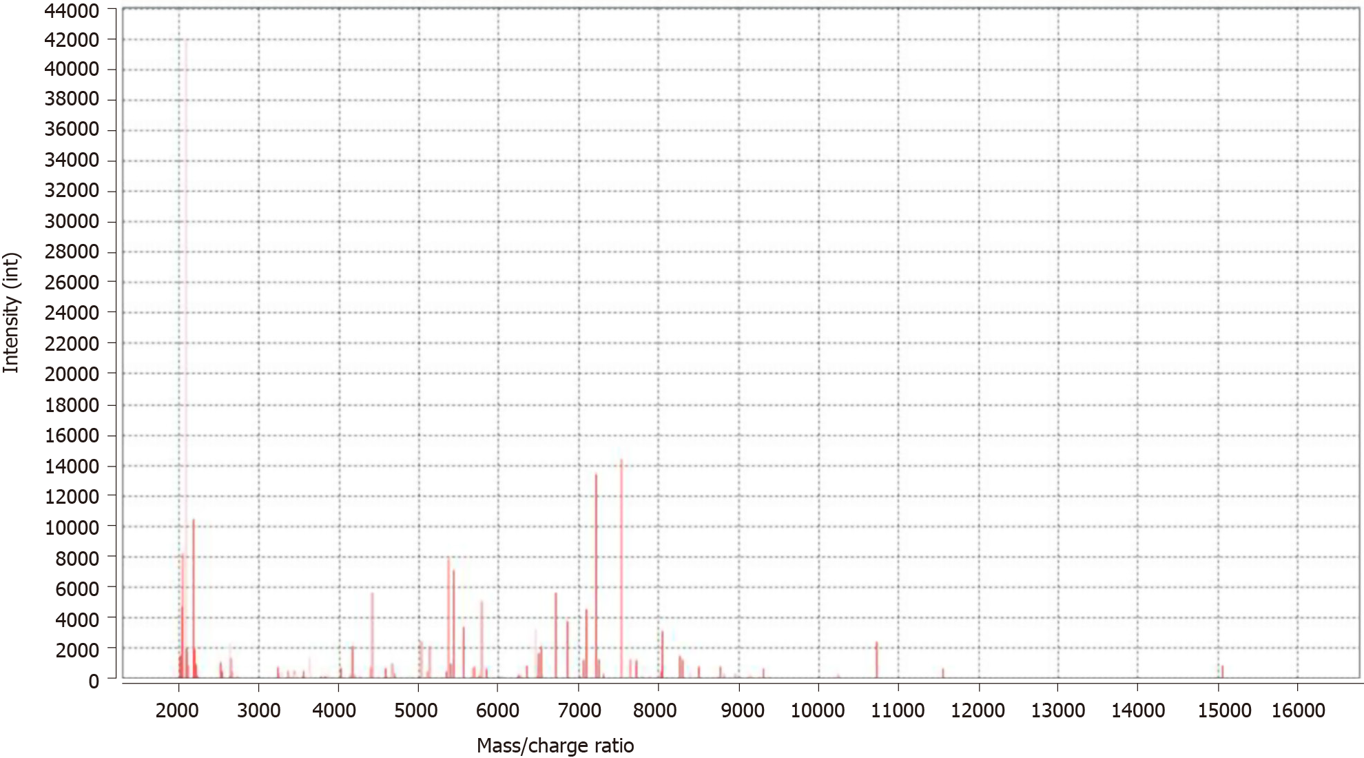
Figure 4 Bacterial mass spectrometry.
Nocardia was identified. The horizontal axis represents the mass/charge ratio, and the vertical axis represents the absolute intensity.
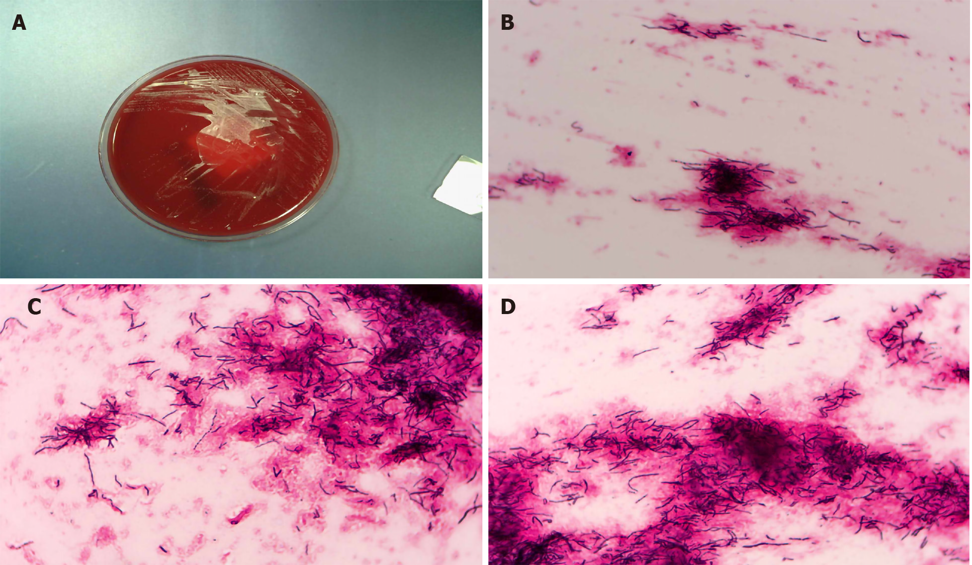
Figure 5 Microscopic appearance of Nocardia in the lung biopsy culture.
A: Bacterial culture colonies; B: Filamentous bacteria were positive for Gram staining (40 ×); C and D: Filamentous bacteria were weakly positive for acid-fast staining (100 ×).
- Citation: Wu XK, Lin Q. Pulmonary alveolar proteinosis complicated with nocardiosis: A case report and review of the literature. World J Clin Cases 2021; 9(12): 2874-2883
- URL: https://www.wjgnet.com/2307-8960/full/v9/i12/2874.htm
- DOI: https://dx.doi.org/10.12998/wjcc.v9.i12.2874


