Copyright
©The Author(s) 2021.
World J Clin Cases. Apr 26, 2021; 9(12): 2862-2867
Published online Apr 26, 2021. doi: 10.12998/wjcc.v9.i12.2862
Published online Apr 26, 2021. doi: 10.12998/wjcc.v9.i12.2862
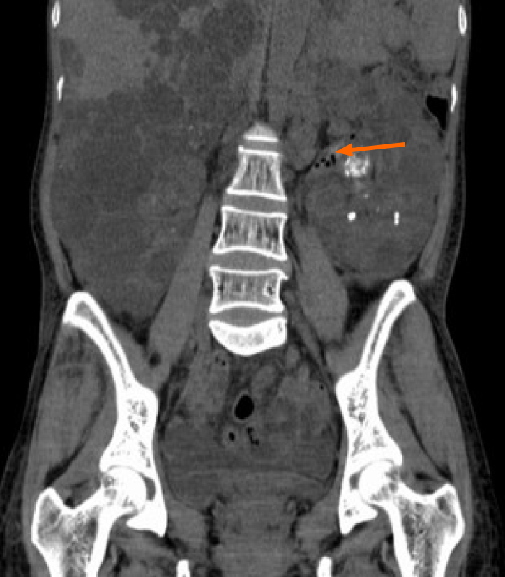
Figure 1
Computed tomography revealed left emphysematous pyelonephritis and multiple renal stones with autosomal dominant polycystic kidney disease.
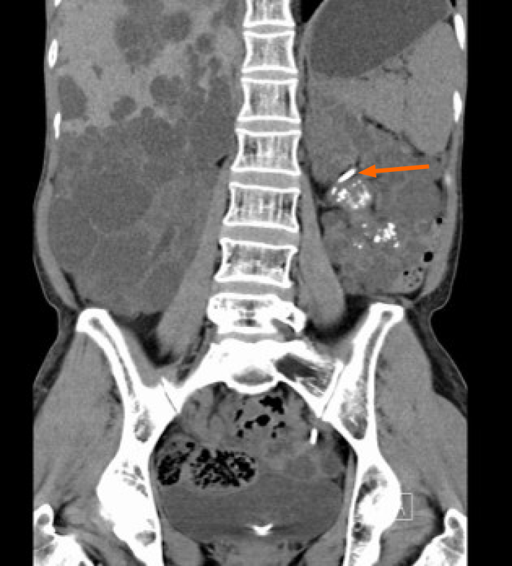
Figure 2 Postoperative computed tomography image.
Postoperative computed tomography showed that the left D-J tube was well positioned, the air spaces in the left collecting system had completely disappeared, and the left hydronephrosis was significantly better than before.
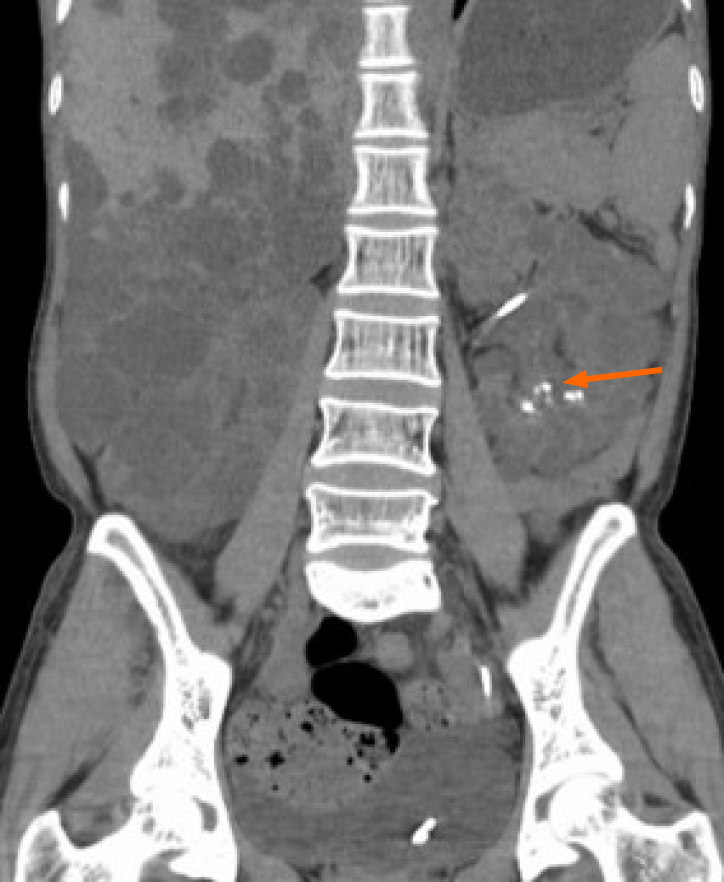
Figure 3 Image after 2nd surgery computed tomography.
Computed tomography after 2nd surgery showed that the stones in the left renal pelvis had been cleared, while just a few stones remained in the lower calyx.
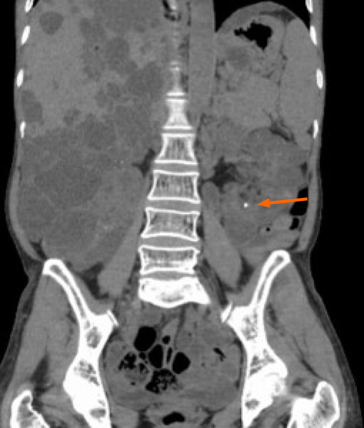
Figure 4 Computed tomography image.
Computed tomography showed no stones in the left renal pelvis, and the stones in the lower calyx were also significantly smaller in size and fewer in number than before.
- Citation: Jiang Y, Lo R, Lu ZQ, Cheng XB, Xiong L, Luo BF. Successful endoscopic surgery for emphysematous pyelonephritis in a non-diabetic patient with autosomal dominant polycystic kidney disease: A case report. World J Clin Cases 2021; 9(12): 2862-2867
- URL: https://www.wjgnet.com/2307-8960/full/v9/i12/2862.htm
- DOI: https://dx.doi.org/10.12998/wjcc.v9.i12.2862


