Copyright
©The Author(s) 2021.
World J Clin Cases. Apr 26, 2021; 9(12): 2845-2853
Published online Apr 26, 2021. doi: 10.12998/wjcc.v9.i12.2845
Published online Apr 26, 2021. doi: 10.12998/wjcc.v9.i12.2845
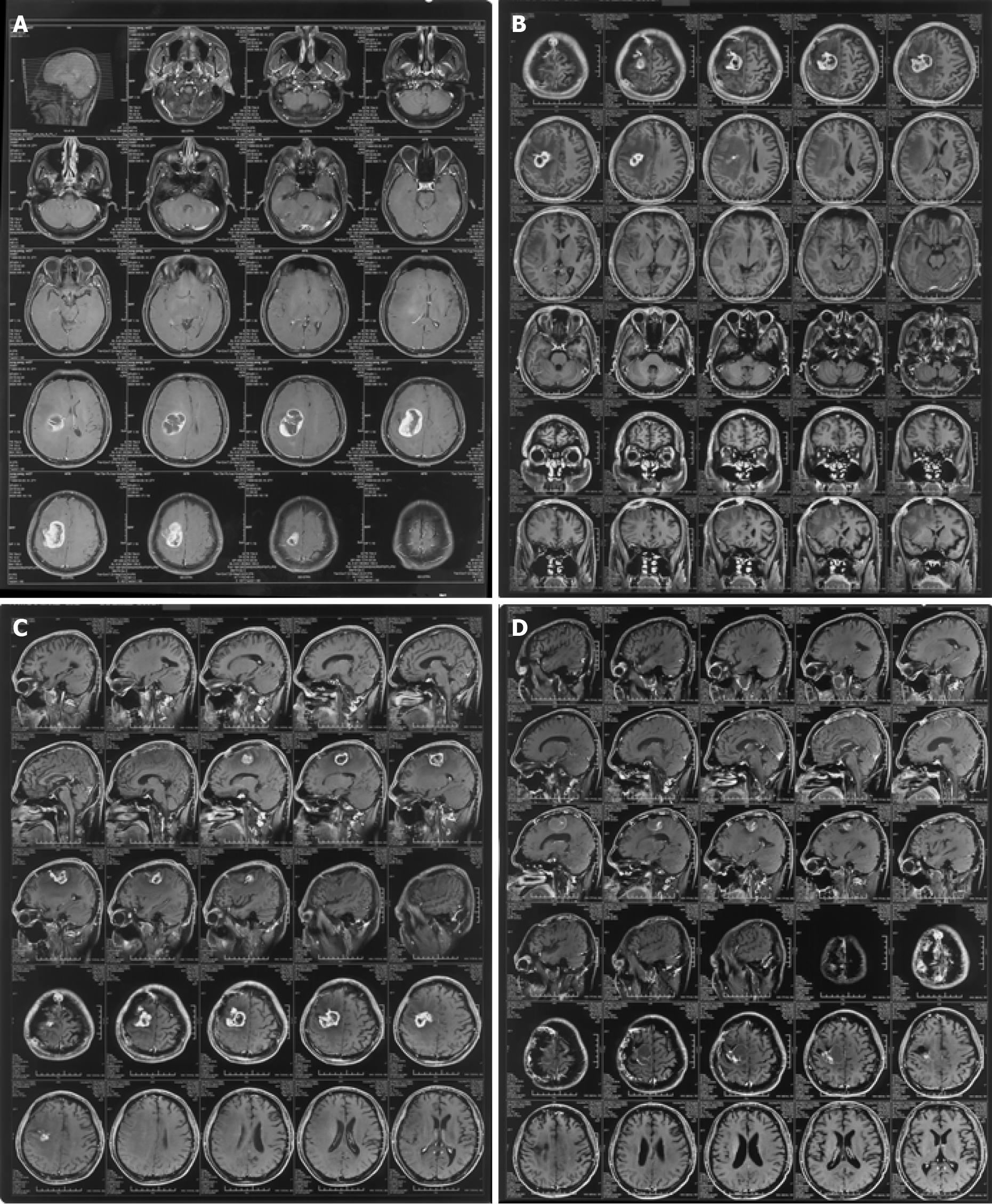
Figure 1 Neuroimaging findings.
A: February 20, 2016 Preoperative image of enhanced right frontal parietal lobe space-occupying lesion, maximum diameter about 5 cm; B: June 3, 2016 End of chemoradiation. Enhanced right frontal parietal lobe space-occupying lesion, maximum diameter about 3.5 cm; C: September 14, 2016 Enhanced lesion at the junction of the right frontal and parietal lobes, maximum diameter about 2 cm, after taking Kangliu pill for 3 mo; and D: April 29, 2019 Enhanced lesion at the junction of the right frontal and parietal lobes, maximum diameter about 1 cm, after taking Kangliu pill for nearly 3 years.
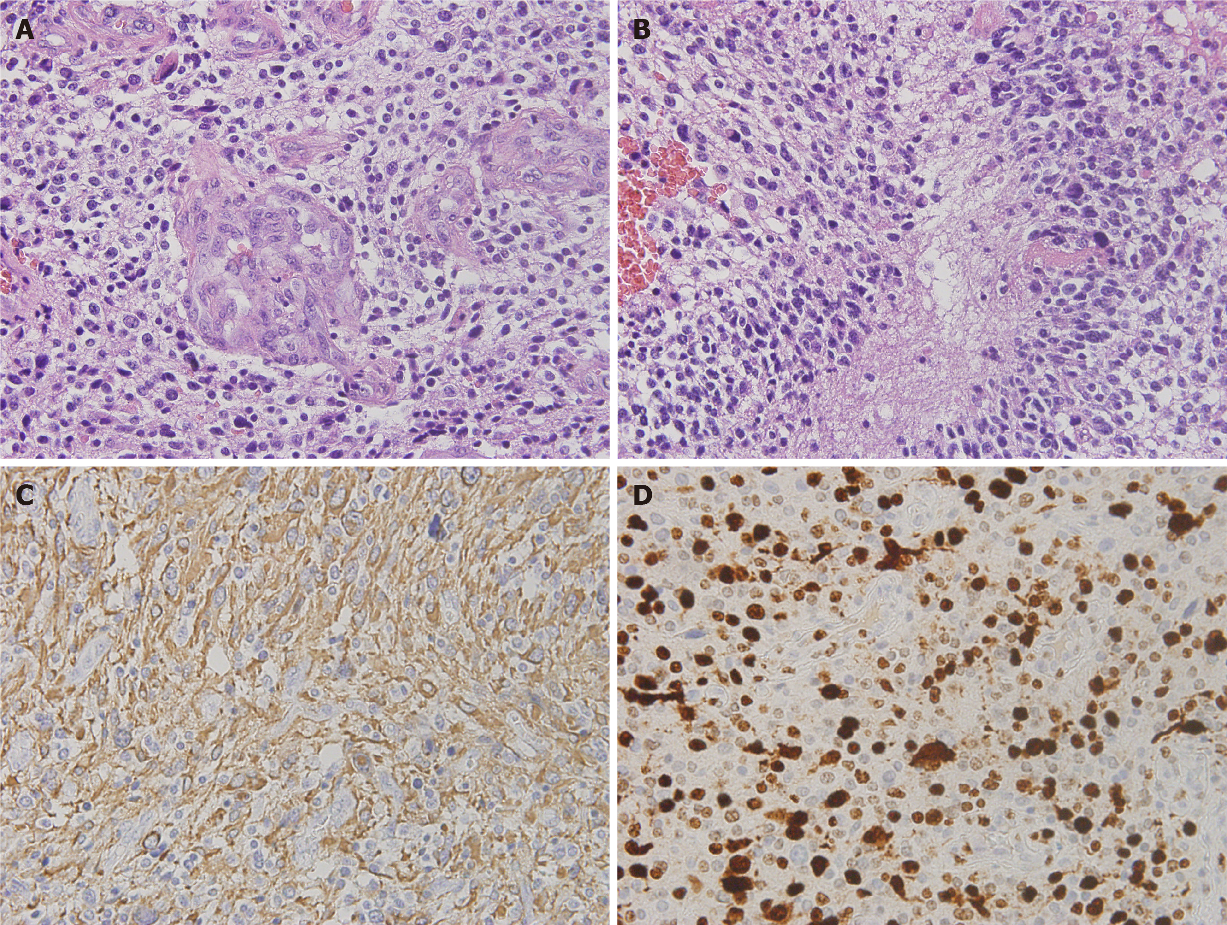
Figure 2 Pathological findings.
A: Hematoxylin-eosin staining showed that the tumor cells diffused and infiltrated; B: Cells were heteromorphic with mitotic figures, microvessel hyperplasia, and palisade necrosis. Original magnification, × 40; C: Tumor cells were immunopositive for glial fibrillary acidic protein immunostaining. Original magnification, × 40; and D: About 70% of tumor cells were positive for Ki-67. Original magnification, × 40.
- Citation: Sun G, Zhuang W, Lin QT, Wang LM, Zhen YH, Xi SY, Lin XL. Partial response to Chinese patent medicine Kangliu pill for adult glioblastoma: A case report and review of the literature. World J Clin Cases 2021; 9(12): 2845-2853
- URL: https://www.wjgnet.com/2307-8960/full/v9/i12/2845.htm
- DOI: https://dx.doi.org/10.12998/wjcc.v9.i12.2845


