Copyright
©The Author(s) 2021.
World J Clin Cases. Apr 26, 2021; 9(12): 2801-2810
Published online Apr 26, 2021. doi: 10.12998/wjcc.v9.i12.2801
Published online Apr 26, 2021. doi: 10.12998/wjcc.v9.i12.2801
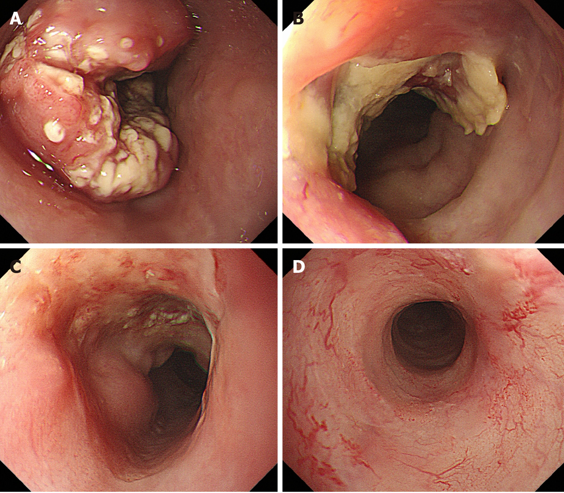
Figure 1 Endoscopic findings.
A: Initial endoscopic findings detected type 2 tumor located on the middle part of the esophagus; B: After 3 courses of induction docetaxel, cisplatin and fluorouracil chemotherapy. Primary tumor volume decreased; C: After subsequent definitive chemoradiotherapy. Esophagogastroduodenoscopy findings showed a prominent reduction of the primary tumor, with ulceration; D: Fourteen months after additional 2 courses cisplatin plus 5fluorouracil chemotherapy. Primary lesion maintained clinically and pathologically complete response.
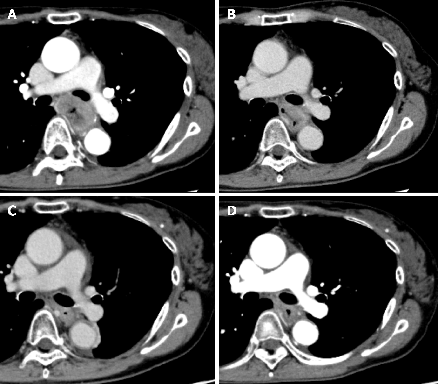
Figure 2 Computed tomography findings showing level of left bronchus.
A: Initial computed tomography findings suspected to invasion to the left bronchus; B: After 3 courses of induction docetaxel, cisplatin and fluorouracil. Shrinkage of the tumor size was recognized but invasion of the left bronchus was still suspected; C: After subsequent definitive chemoradiotherapy. Primary tumor volume decreased; D: Fourteen months after additional 2 courses of cisplatin plus 5fluorouracil chemotherapy. Primary tumor has maintained shrinkage.
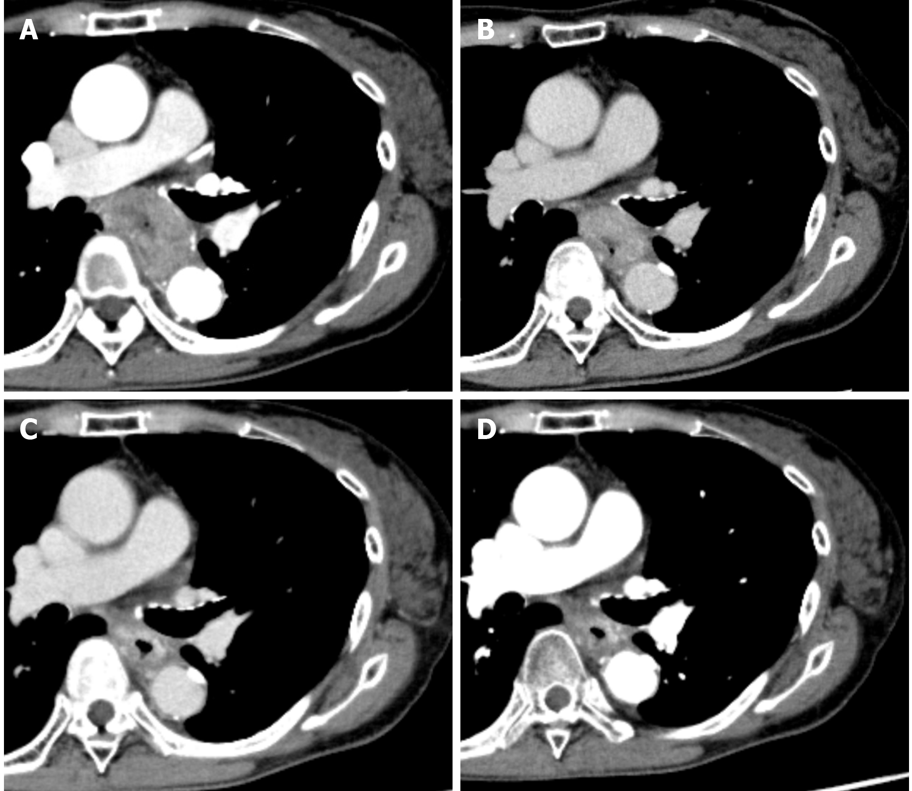
Figure 3 Computerized tomography findings showing level of aorta.
A: Initial computed tomography findings suspected to 90° of direct contact with the aorta, which defined as invasion to the aorta; B: After 3 courses of induction docetaxel, cisplatin and fluorouracil. Shrinkage of the tumor size was recognized but invasion of the aorta was still suspected; C: After subsequent definitive chemoradiotherapy. Primary tumor volume decreased; D: Fourteen months after additional 2 courses of cisplatin plus 5fluorouracil chemotherapy. Primary tumor has maintained shrinkage.
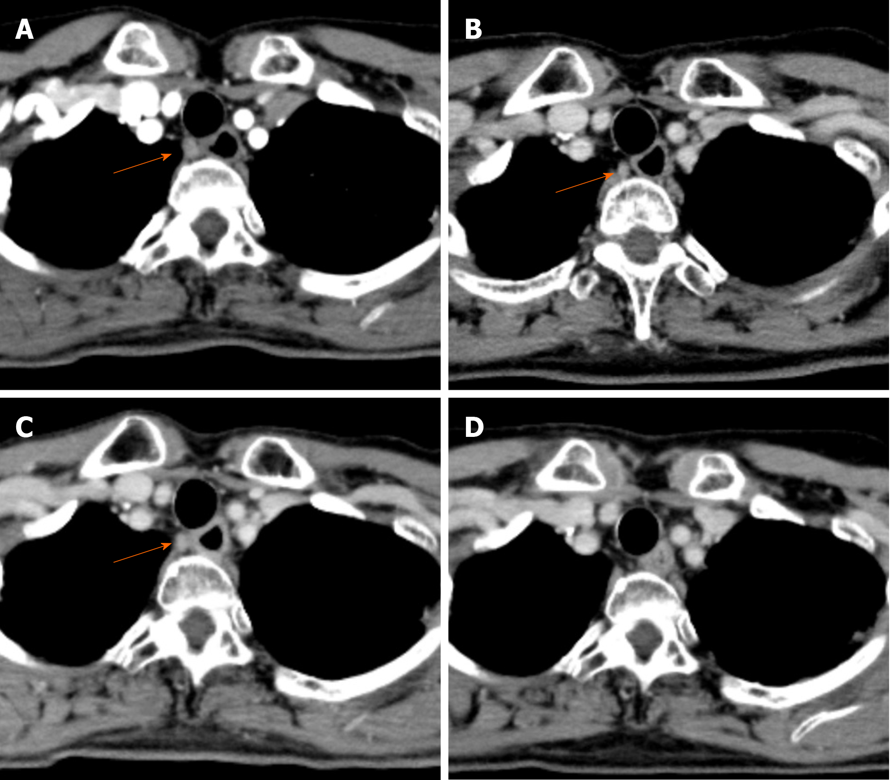
Figure 4 Computed tomography findings showing #106recR lymph node.
A: Initial computed tomography findings suspected swollen lymph node (10.16 mm × 8.02 mm); B: After 3 courses of induction docetaxel, cisplatin and fluorouracil. Slightly shrinkage of the lymph node size (8.01 mm × 6.69 mm) was recognized; C: After subsequent definitive chemoradiotherapy. #106recR lymph node did not change in size (8.31 mm × 7.59 mm); D: After additional 2 courses of cisplatin plus 5fluorouracil chemotherapy. #106recR lymph node could not be detected (Orange arrow; 106recR lymph node).
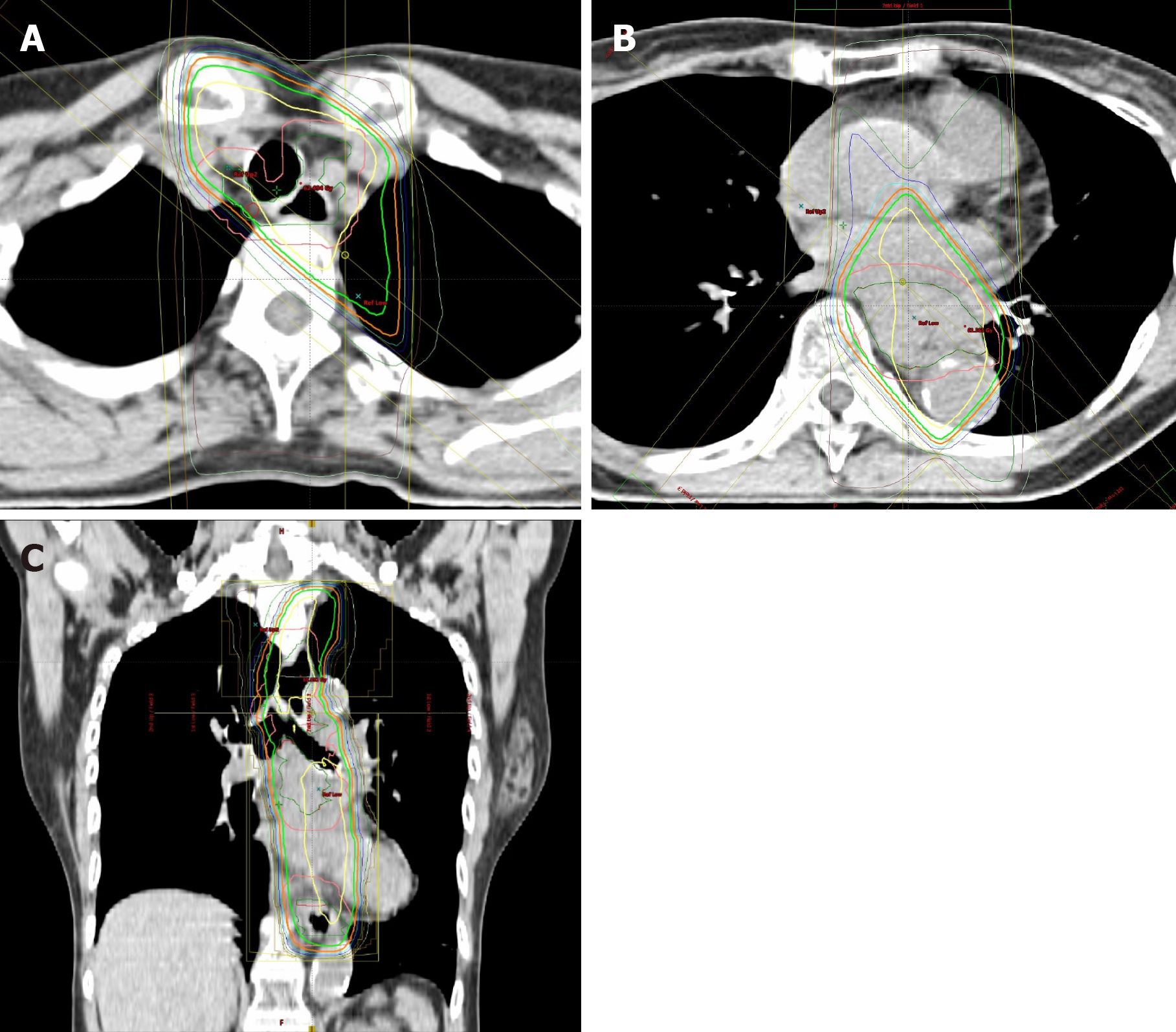
Figure 5 Chemoradiation planning.
A: Swollen lymph node (#106recR) was designated as gross tumor volume node; B and C: The clinical target volume included the gross tumor volume and a craniocaudal margin of 2.4 cm around the primary tumor.
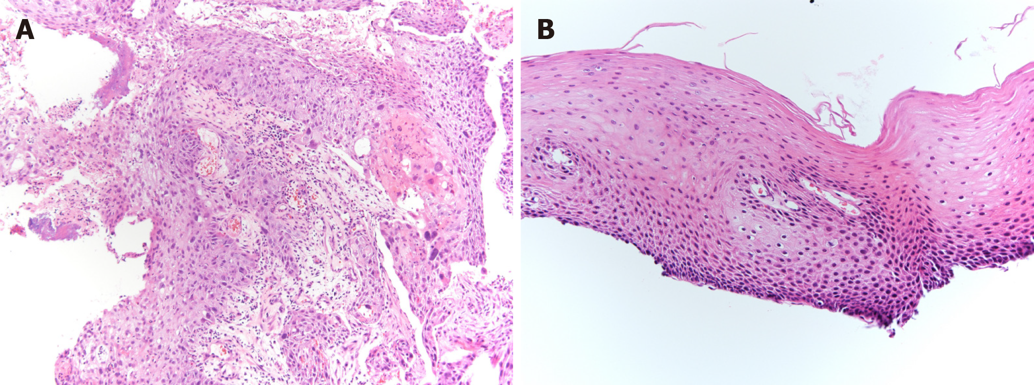
Figure 6 Pathological examination, biopsy from primary lesion.
A: Initial biopsy from primary tumor revealed squamous cell carcinoma (× 10). A proliferation of large atypical cells with swollen deep-stained nuclei was observed. A squamous cell carcinoma with a tendency toward keratinization, intercellular bridges, necrosis, and large atypical cells with bizarre nuclei were evident; B: Fourteen months after additional 2 courses of cisplatin plus 5fluorouracil chemotherapy, primary lesion has maintained pathologically negative for cancer (× 20). There is a small infiltration of inflammatory cells, but no obvious evidence of malignant cells.
- Citation: Yura M, Koyanagi K, Hara A, Hayashi K, Tajima Y, Kaneko Y, Fujisaki H, Hirata A, Takano K, Hongo K, Yo K, Yoneyama K, Tamai Y, Dehari R, Nakagawa M. Unresectable esophageal cancer treated with multiple chemotherapies in combination with chemoradiotherapy: A case report. World J Clin Cases 2021; 9(12): 2801-2810
- URL: https://www.wjgnet.com/2307-8960/full/v9/i12/2801.htm
- DOI: https://dx.doi.org/10.12998/wjcc.v9.i12.2801


