Copyright
©The Author(s) 2021.
World J Clin Cases. Apr 26, 2021; 9(12): 2751-2762
Published online Apr 26, 2021. doi: 10.12998/wjcc.v9.i12.2751
Published online Apr 26, 2021. doi: 10.12998/wjcc.v9.i12.2751
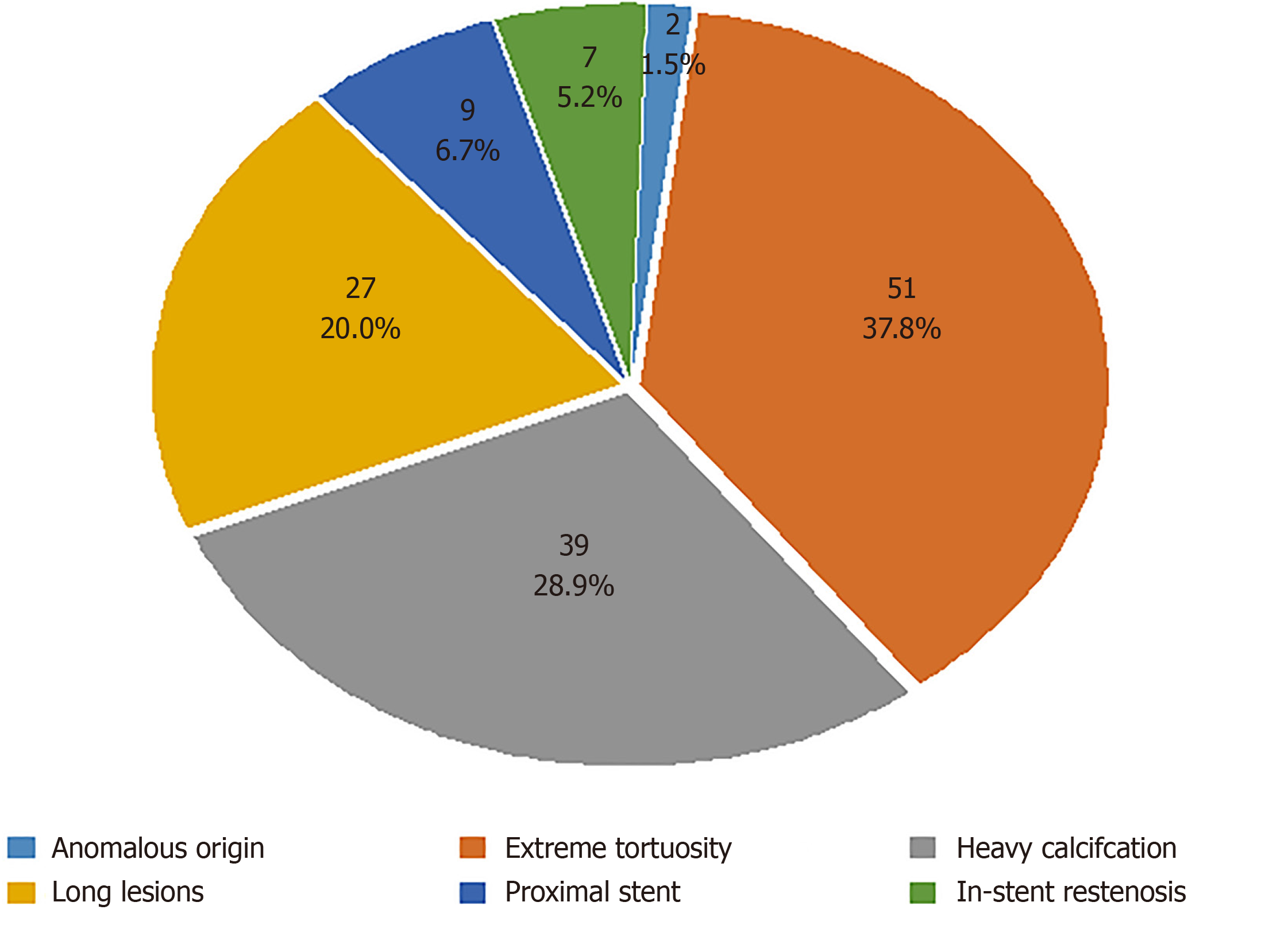
Figure 1 Target lesion characteristics.
Figure 2 A patient with coronary artery origin anomaly.
A: The right coronary artery originated from the left coronary sinus. The left anterior descending artery and left circumflex artery were visible during right coronary artery angiography; B: The stent was successfully passed through the lesion and implanted with the aid of a rapid exchange extension catheter; C: Final angiographic result.
Figure 3 An extremely tortuous lesion of the distal segment of the right coronary artery.
A: A lesion located in the distal segment of right coronary artery before the bifurcation, with extreme tortuosity in the proximal and middle segments of the right coronary artery; B: A stent (2.5 mm × 13 mm) was implanted with the use of the rapid exchange extension catheter; C: Final angiographic result.
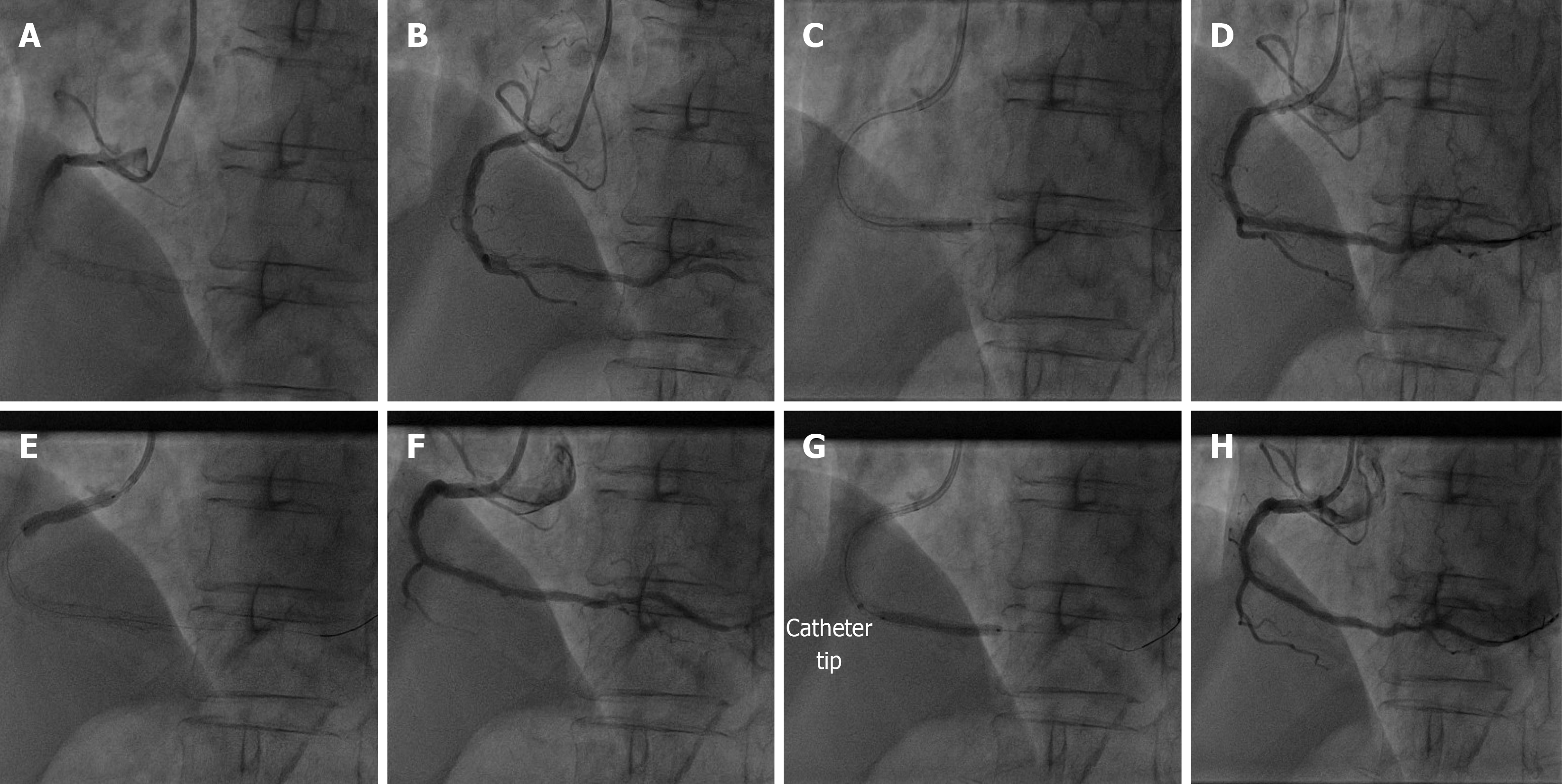
Figure 4 A case with in-stent restenosis was treated with drug-coated balloon.
A: The stent was visible in the middle-distal segment of the right coronary artery (RCA); B: Angiography revealed in-stent restenosis; C: A balloon predilated the in-stent restenosis lesion; D: Angiography after predilatation showed severe stenosis and tortuosity in proximal RCA; E: Preimplantation of a stent in the proximal segment of the RCA; F: Angiography after stenting the proximal RCA; G: An in-stent restenosis lesion was treated with drug-coated balloon using a rapid exchange extension catheter; H: Final angiographic result.
- Citation: Wang HC, Lu W, Gao ZH, Xie YN, Hao J, Liu JM. Application of a rapid exchange extension catheter technique in type B2/C nonocclusive coronary intervention via a transradial approach. World J Clin Cases 2021; 9(12): 2751-2762
- URL: https://www.wjgnet.com/2307-8960/full/v9/i12/2751.htm
- DOI: https://dx.doi.org/10.12998/wjcc.v9.i12.2751


