Copyright
©The Author(s) 2021.
World J Clin Cases. Apr 26, 2021; 9(12): 2739-2750
Published online Apr 26, 2021. doi: 10.12998/wjcc.v9.i12.2739
Published online Apr 26, 2021. doi: 10.12998/wjcc.v9.i12.2739
Figure 1 Computed tomography showed that the tumor was located in the space between the sartorius muscle and tensor fascia lata muscle in front of the right thigh.
A: Plain computed tomography scan of different layers of case 3 revealed that the tumor was located in the subcutaneous adipose tissue, and was well-circumscribed; B: Enhanced computed tomography revealed diffuse and weak enhancement in the tumor. Blue arrows indicate the tumor.
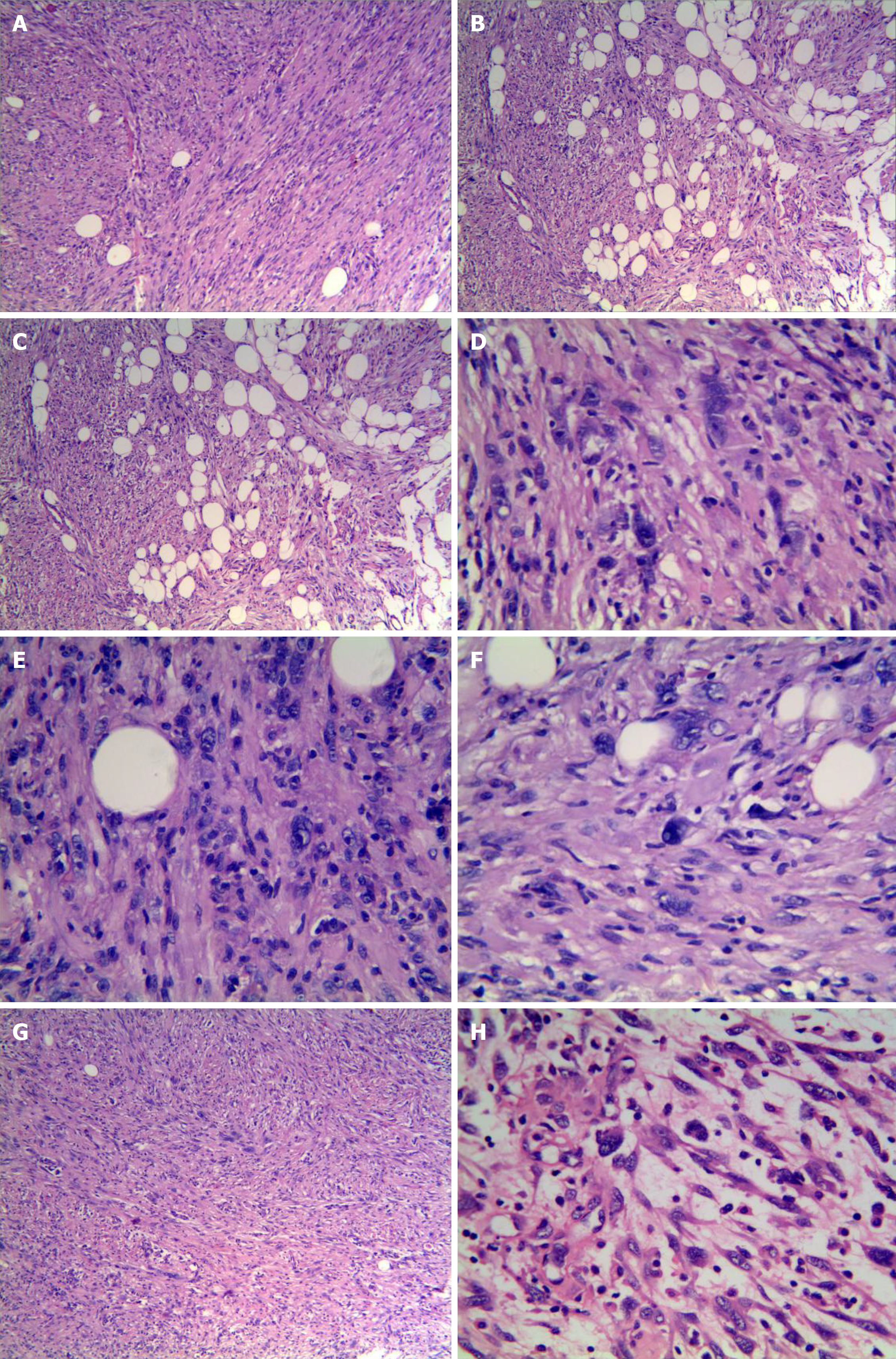
Figure 2 Histological features of the tumor stained with hematoxylin and eosin.
A: Relatively monomorphic spindled cells growing in intersecting fascicles (magnification, × 100); B: Infiltrating into the surrounding fibroadipose tissue focally (magnification, × 100); C: Epithelioid cells in the local area arranged in sheets and nests(magnification, × 400); D-F: A few scattered tumors cells exhibited irregularly hyperchromatic, bizarre, and pleomorphic nuclei with frequent intranuclear pseudoinclusions (black arrows) and rare mitotic figures(magnification, × 400); G: In some areas, tumor morphology was similar to that of inflammatory myofibroblastoma (magnification, × 100); H: In other areas, tumor morphology was similar to that of myxoinflammatory fibroblastosarcoma (magnification, × 400).
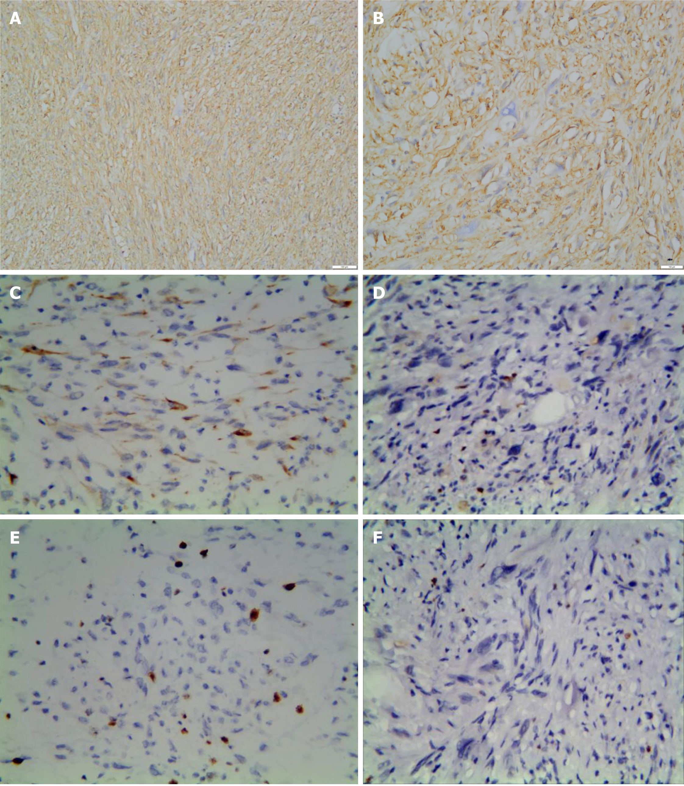
Figure 3 Immunohistochemica characteristics of the tumor.
A and B: CD34 was strongly and diffusely expressed in almost all of the tumor cells (magnification, A: × 100; B: × 400); C: Two cases were focally positive for CKpan (magnification, × 100); D: Ki-67 proliferation index was less than 2% (magnification, × 400); E: The proliferative index of case 3 was up to 5% locally (magnification, × 100); F: p53 was negative for tumor cells (magnification, × 400).
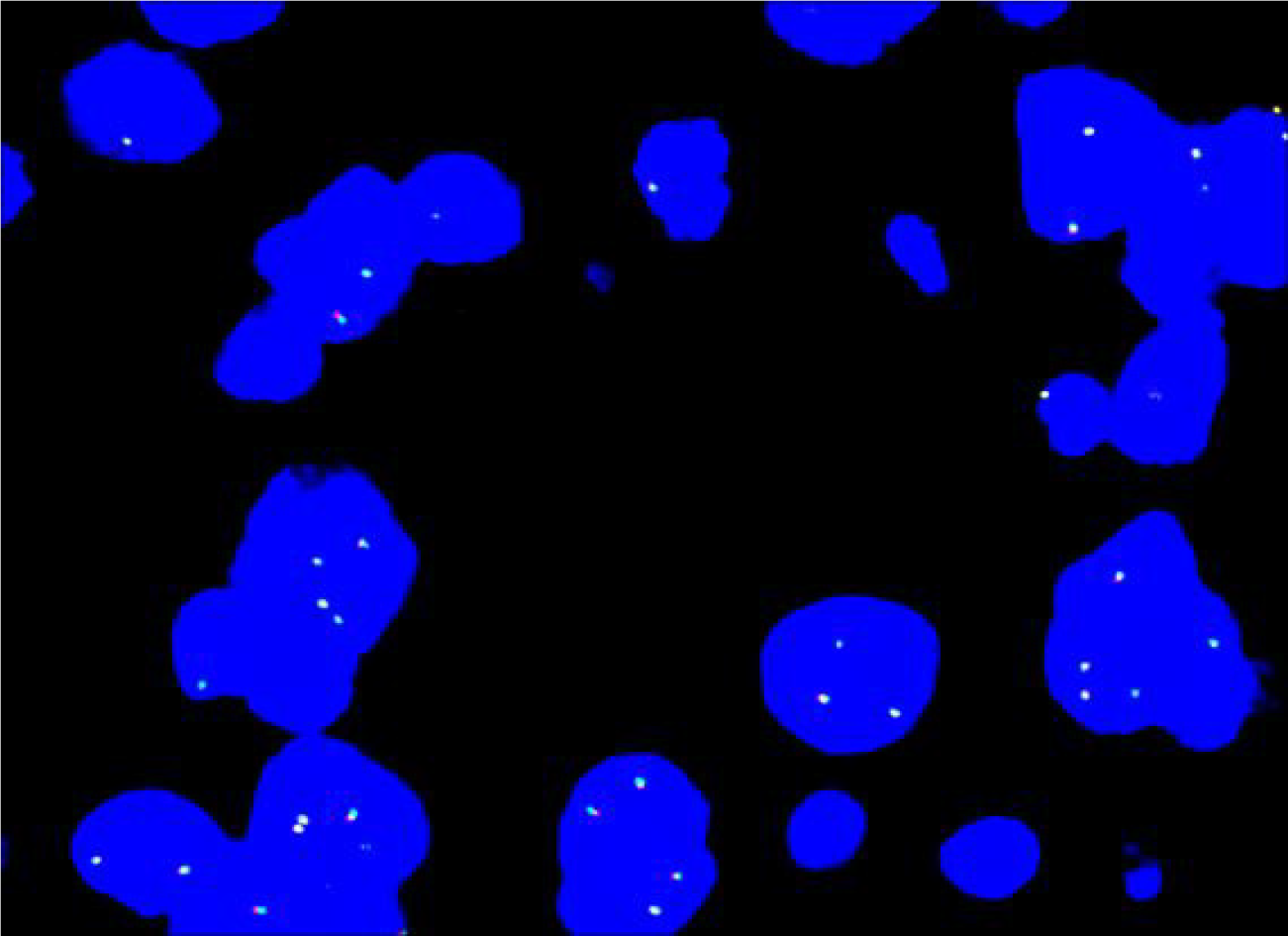
Figure 4 ALK dual-color break-apart rearrangement probe detection by fluorescence in situ hybridization.
All tested cases were negative for the inflammatory myofibroblastoma-associated ALK gene rearrangement.
- Citation: Ding L, Xu WJ, Tao XY, Zhang L, Cai ZG. Clinicopathological features of superficial CD34-positive fibroblastic tumor. World J Clin Cases 2021; 9(12): 2739-2750
- URL: https://www.wjgnet.com/2307-8960/full/v9/i12/2739.htm
- DOI: https://dx.doi.org/10.12998/wjcc.v9.i12.2739


