Copyright
©The Author(s) 2020.
World J Clin Cases. Apr 26, 2020; 8(8): 1525-1531
Published online Apr 26, 2020. doi: 10.12998/wjcc.v8.i8.1525
Published online Apr 26, 2020. doi: 10.12998/wjcc.v8.i8.1525
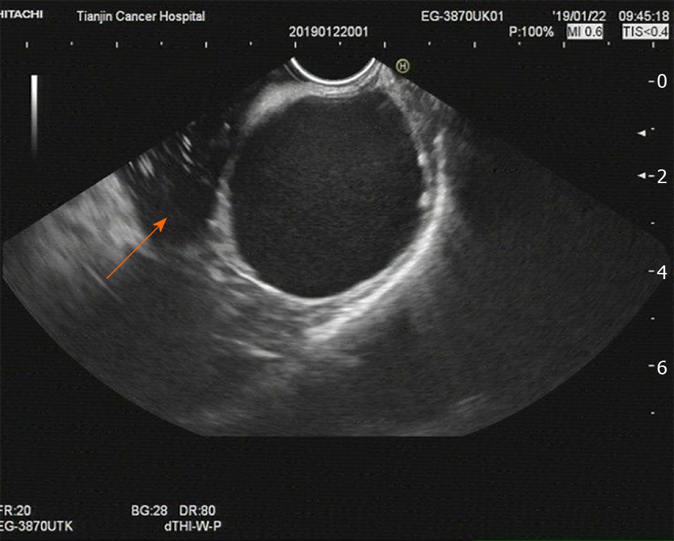
Figure 1 Imaging at admission.
An endoscopic ultrasound examination showed a 45 mm × 49 mm single cyst originating from the gastric submucosa (arrow). It also revealed that the cyst, without any echoes or color flow signals, was located in the posterior wall of the fundus close to the cardia. These results indicated the possibility of cystic hygroma of the stomach.
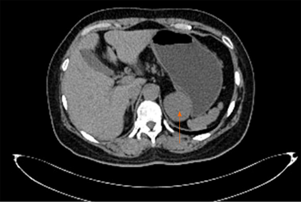
Figure 2 Imaging at admission.
A plain computed tomography scan demonstrated a 52 mm × 43 mm quasi-circular mass (arrow). It appeared to have a low density of 29 HU and was closely related to the lesser curvature of the gastric fundus, which also showed an extraluminal growth pattern with an obvious border.
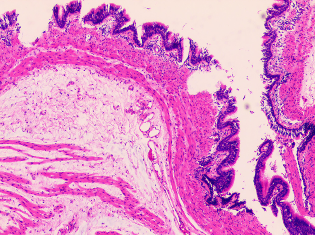
Figure 3 Postoperative histopathology.
Microscopically, pseudostratified ciliated columnar epithelial cells could be observed in the cyst wall, and the structure of these cells was the same as that of the bronchus. Combined with immunohistochemical staining for CK7 (+), CK20 (-), TTF1 (-), and S-100 (-), the pathologists ultimately verified that this cystic mass of the fundus was a gastric bronchogenic cyst (×100).
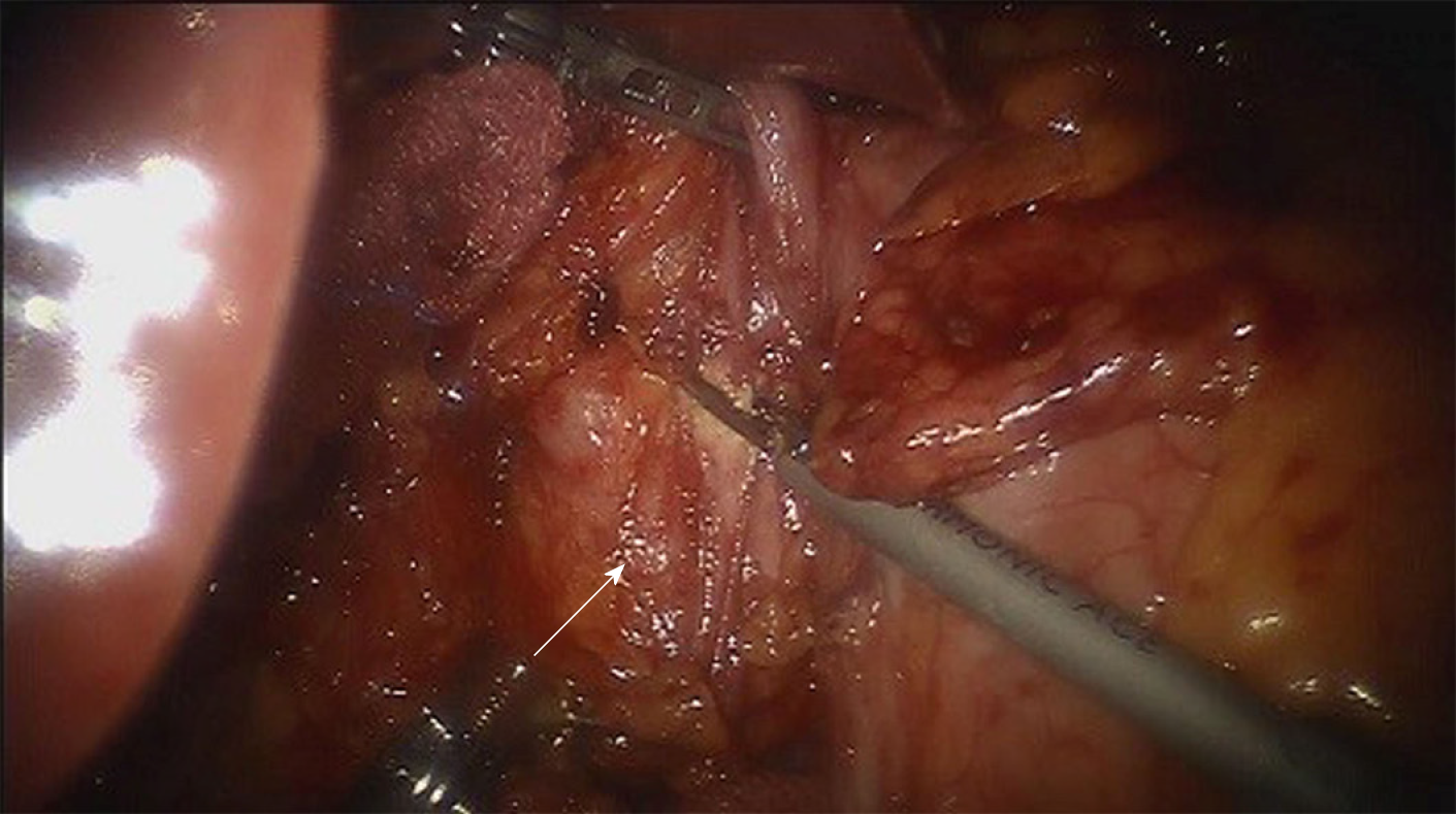
Figure 4 Imaging during operation.
Intraoperative findings of this cystic mass showed that it was a smooth single-port cyst originating from the submucosa of the posterior gastric wall close to the cardia (arrow). Then, the cystic mass was curatively resected from the stomach, and the surgeons paid attention to avoiding cyst rupture simultaneously.
- Citation: He WT, Deng JY, Liang H, Xiao JY, Cao FL. Bronchogenic cyst of the stomach: A case report. World J Clin Cases 2020; 8(8): 1525-1531
- URL: https://www.wjgnet.com/2307-8960/full/v8/i8/1525.htm
- DOI: https://dx.doi.org/10.12998/wjcc.v8.i8.1525


