Copyright
©The Author(s) 2020.
World J Clin Cases. Apr 26, 2020; 8(8): 1489-1494
Published online Apr 26, 2020. doi: 10.12998/wjcc.v8.i8.1489
Published online Apr 26, 2020. doi: 10.12998/wjcc.v8.i8.1489
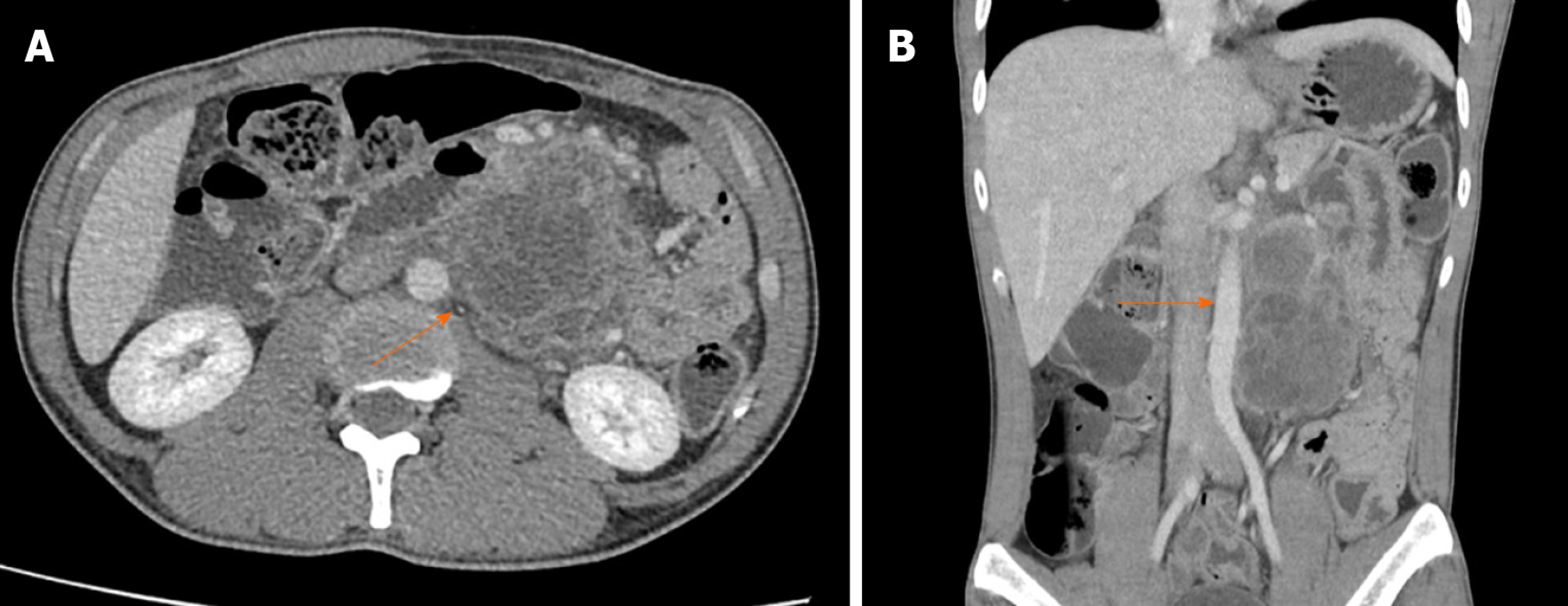
Figure 1 Computed tomography scan of the abdomen.
A: Axial enhanced computed tomography image demonstrated a multiseptated cystic tumor (arrow) in the retroperitoneal region, adhering to the duodenum and close to the abdominal aorta and left renal vein; B: coronal enhanced computed tomography reconstruction demonstrating a tumor close to the abdominal aorta (arrow).
Figure 2 Histopathological findings (hematoxylin-eosin staining, magnification ×100).
A: Teratoma located between the inner circular and the outer longitudinal layers of the duodenal muscularis propria (arrow) and communicate with the normal duodenal mucosa (arrowhead); B: the tumor consists of stratified squamous cell epithelium, simple cuboidal epithelium, and irregular bundles of smooth muscle, fat and hyaline cartilage.
- Citation: Chansoon T, Angkathunyakul N, Aroonroch R, Jirasiritham J. Duodenal mature teratoma causing partial intestinal obstruction: A first case report in an adult. World J Clin Cases 2020; 8(8): 1489-1494
- URL: https://www.wjgnet.com/2307-8960/full/v8/i8/1489.htm
- DOI: https://dx.doi.org/10.12998/wjcc.v8.i8.1489


