Copyright
©The Author(s) 2020.
World J Clin Cases. Apr 26, 2020; 8(8): 1454-1462
Published online Apr 26, 2020. doi: 10.12998/wjcc.v8.i8.1454
Published online Apr 26, 2020. doi: 10.12998/wjcc.v8.i8.1454
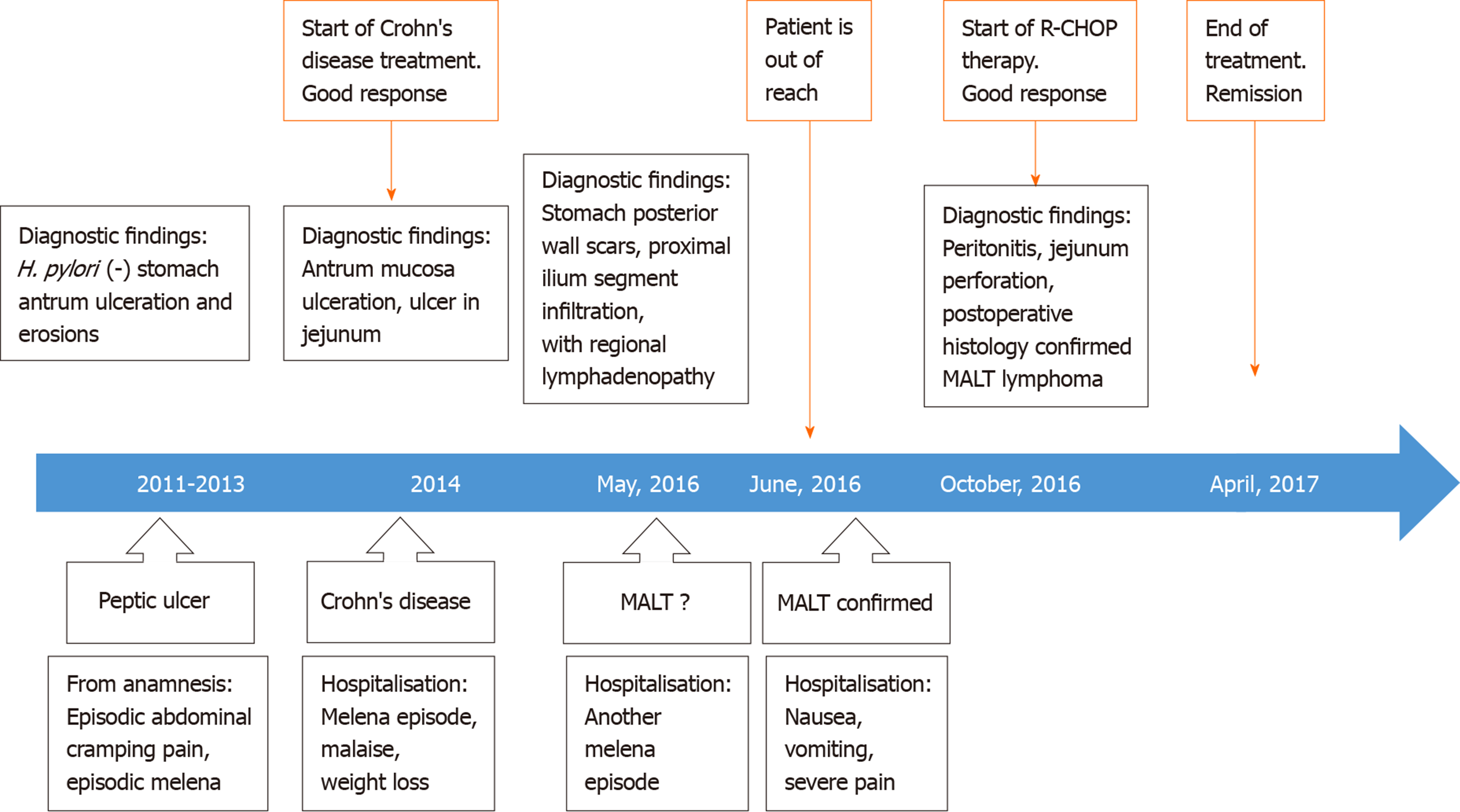
Figure 1 Case history timeline.
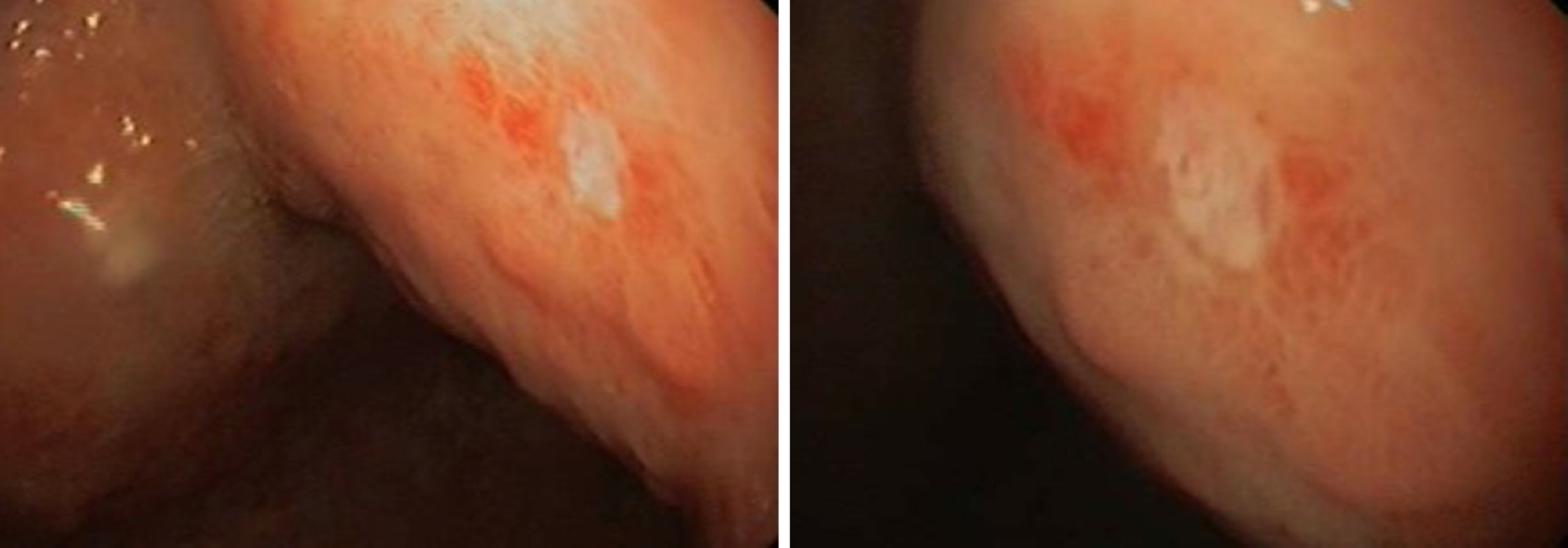
Figure 2 Superficial ulcer in stomach found during upper gastrointestinal endoscopy.
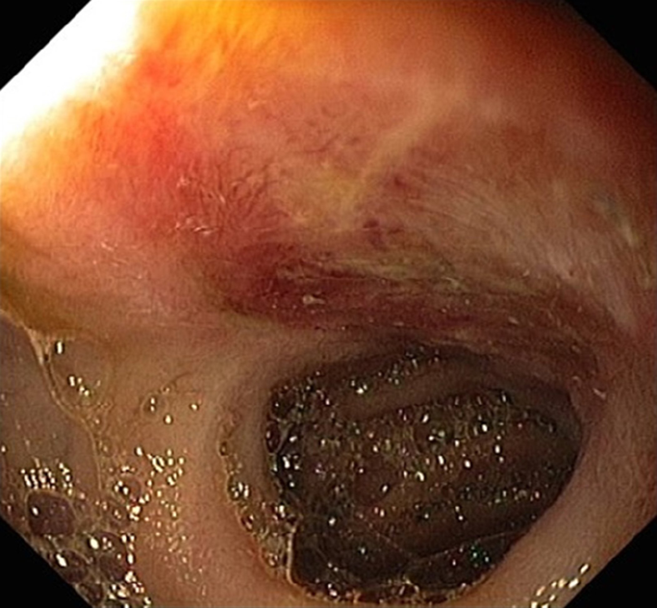
Figure 3 Ulcer in jejunum found during enteroscopy.
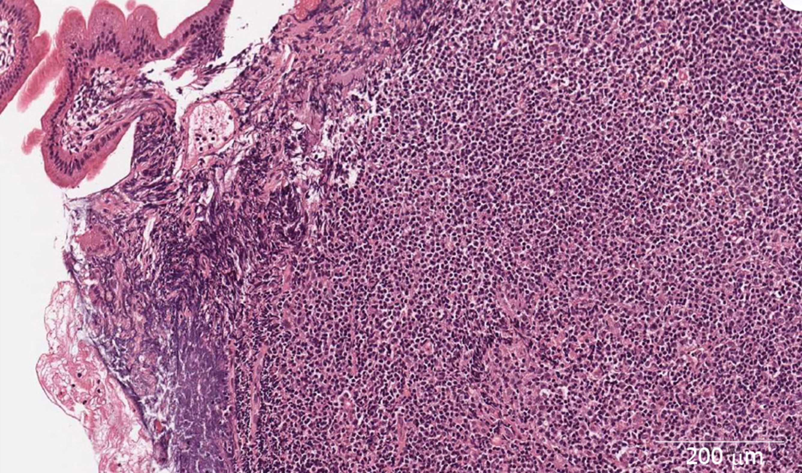
Figure 4 Lymphoplasmacytic infiltrate of jejunum mucosa with eosinophils and neutrophils (× 400).
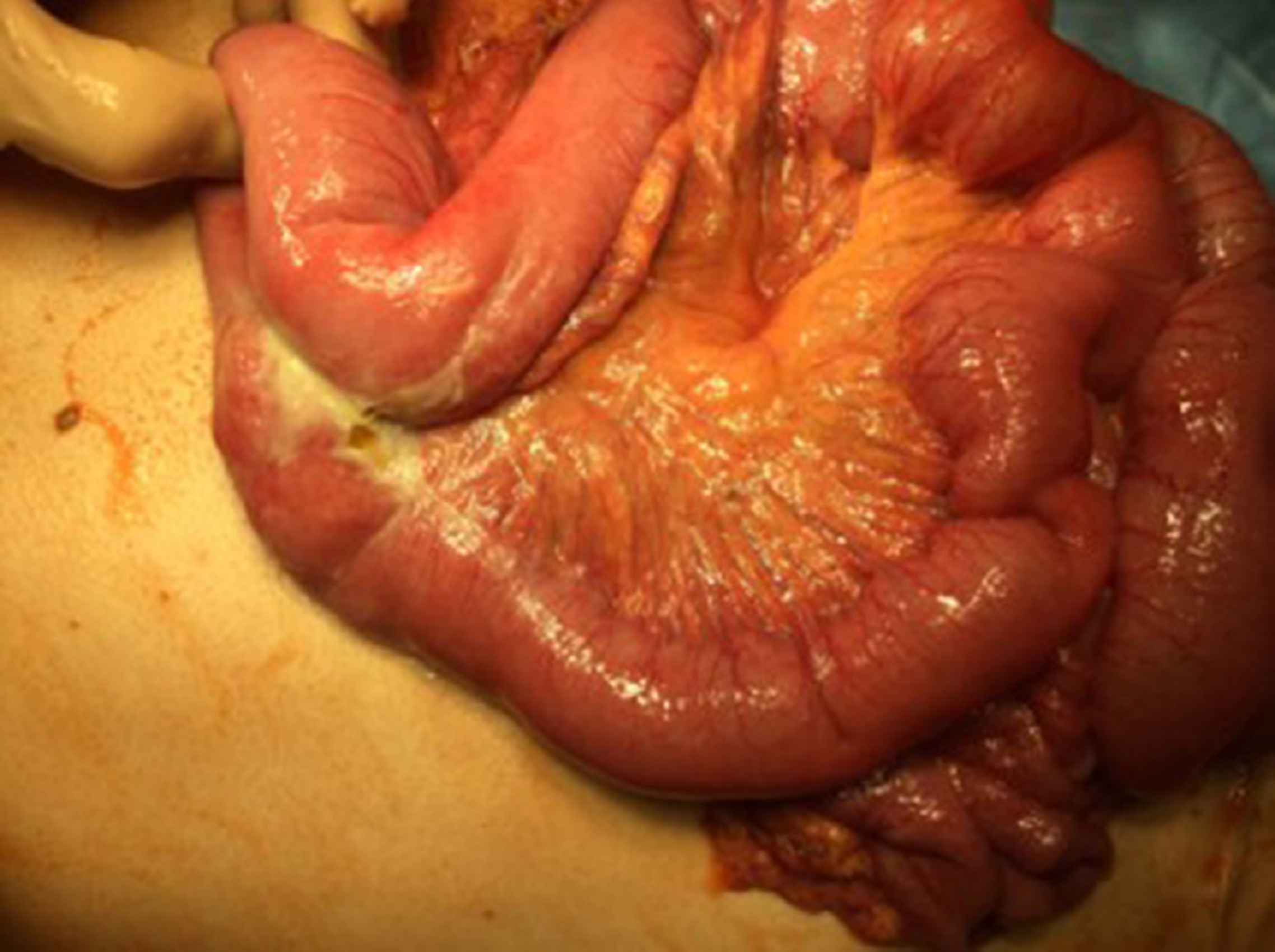
Figure 5 Intra-operative picture of jejunal perforation.
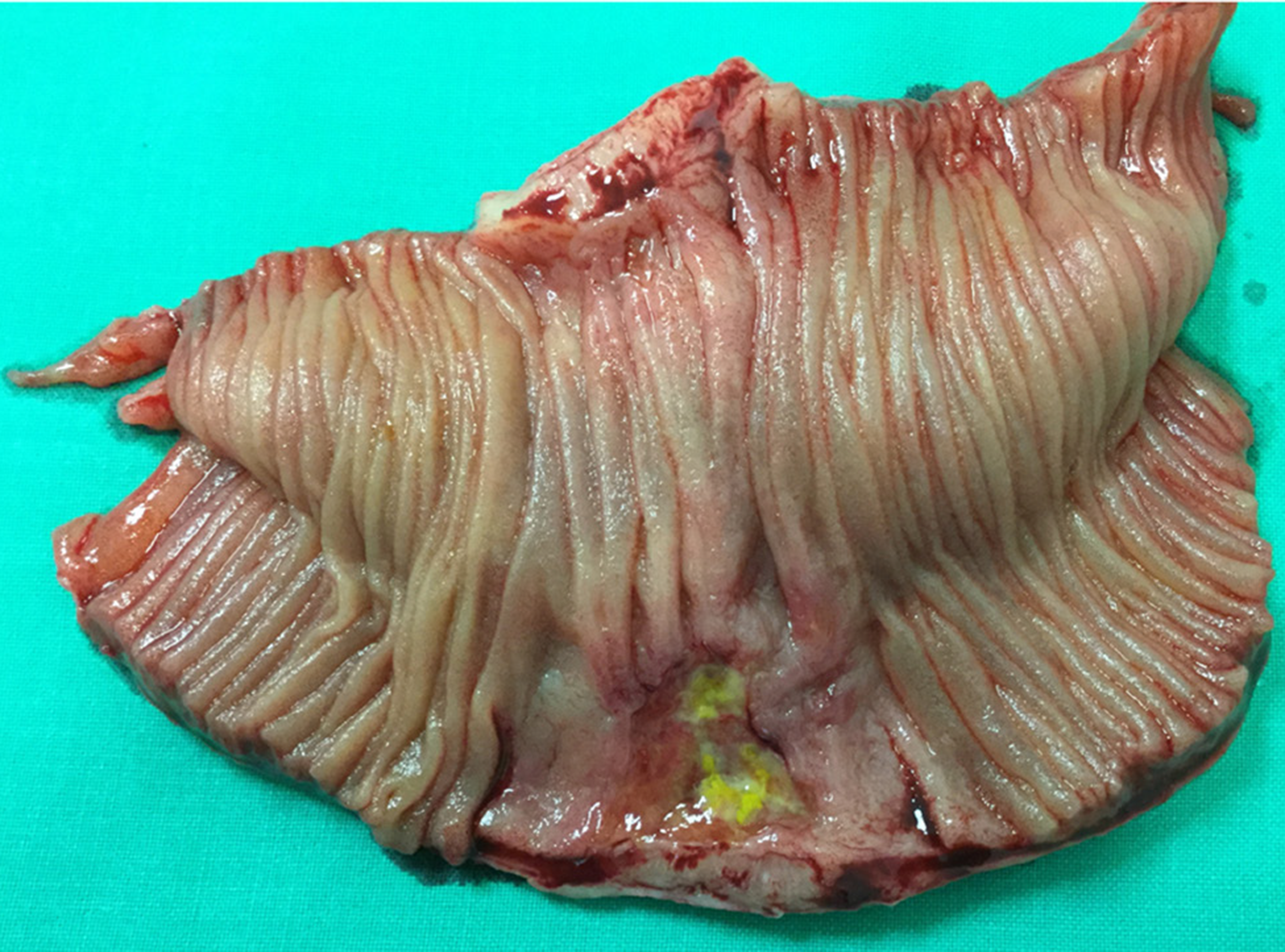
Figure 6 The resected segment of jejunum and mesentery.
Figure 7 Histology of the postoperative jejunum disclosed jejunal mucosa-associated lymphoid tissue lymphoma.
A: Dense population of atypical lymphoid cells in jejunum mucosa (× 200); B: CD20 immunohistochemical staining (× 400).
- Citation: Stundiene I, Maksimaityte V, Liakina V, Valantinas J. Mucosa-associated lymphoid tissue lymphoma simulating Crohn’s disease: A case report. World J Clin Cases 2020; 8(8): 1454-1462
- URL: https://www.wjgnet.com/2307-8960/full/v8/i8/1454.htm
- DOI: https://dx.doi.org/10.12998/wjcc.v8.i8.1454


