Copyright
©The Author(s) 2020.
World J Clin Cases. Apr 6, 2020; 8(7): 1271-1277
Published online Apr 6, 2020. doi: 10.12998/wjcc.v8.i7.1271
Published online Apr 6, 2020. doi: 10.12998/wjcc.v8.i7.1271
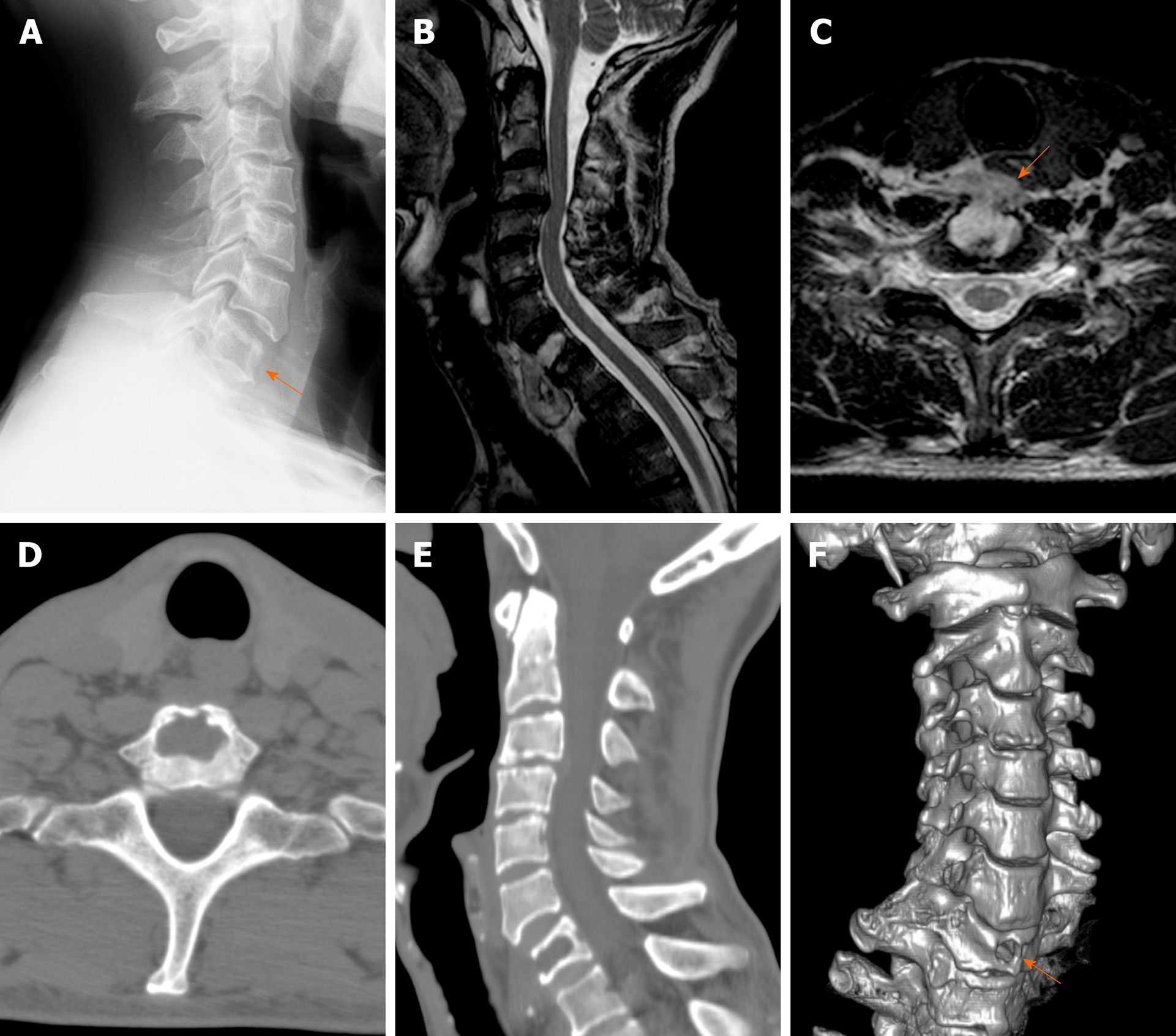
Figure 1 Imaging findings before treatment.
A: Cervical X-ray showed loss of height of the seventh cervical (C7) vertebral body; B: Sagittal T2-weighted magnetic resonance imaging of the cervical spine showing a hyperintense prevertebral mass; C: Axial magnetic resonance imaging showed a well-demarcated lesion seemingly originating in corpus C7 without spinal canal and neuroforamina involvement; D: Axial computed tomography (CT) shows lytic changes in the anterior cortical margin of C7 body. Posterior cortical margin is intact; E: CT scanning exhibited a bony defect of the C7 body; F: Three-dimensional CT reconstruction shows a circular defect in the anterior edge of the C7 vertebra.
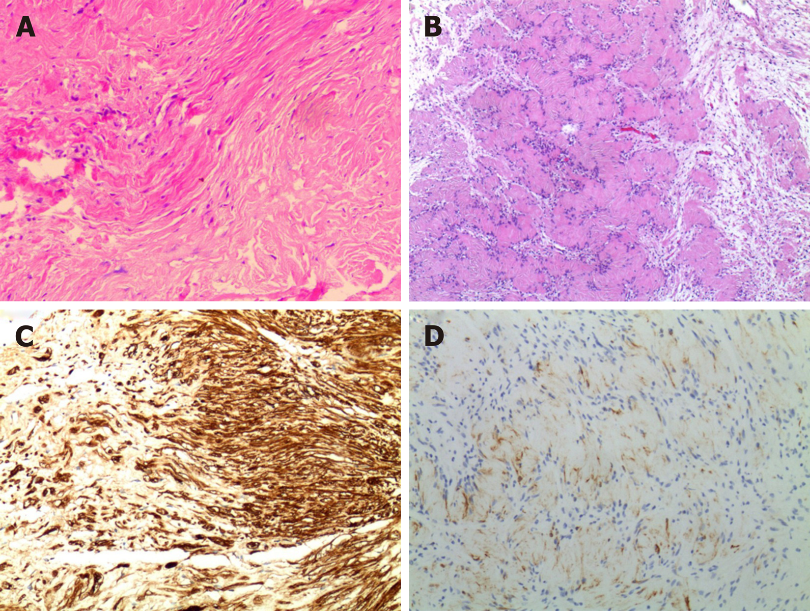
Figure 2 Histological examination of the lesion (hematoxylin and eosin; × 200).
A: Histology revealed hypercellular (Antoni A) and hypocellular (Antoni B) areas, indicating a typical schwannoma; B: Tumor cells constitute the Verocay; C: Immunohistochemical study for S100 protein is diffusely positive; D: Immunohistochemistry showed positive glial fibrillary acidic protein.
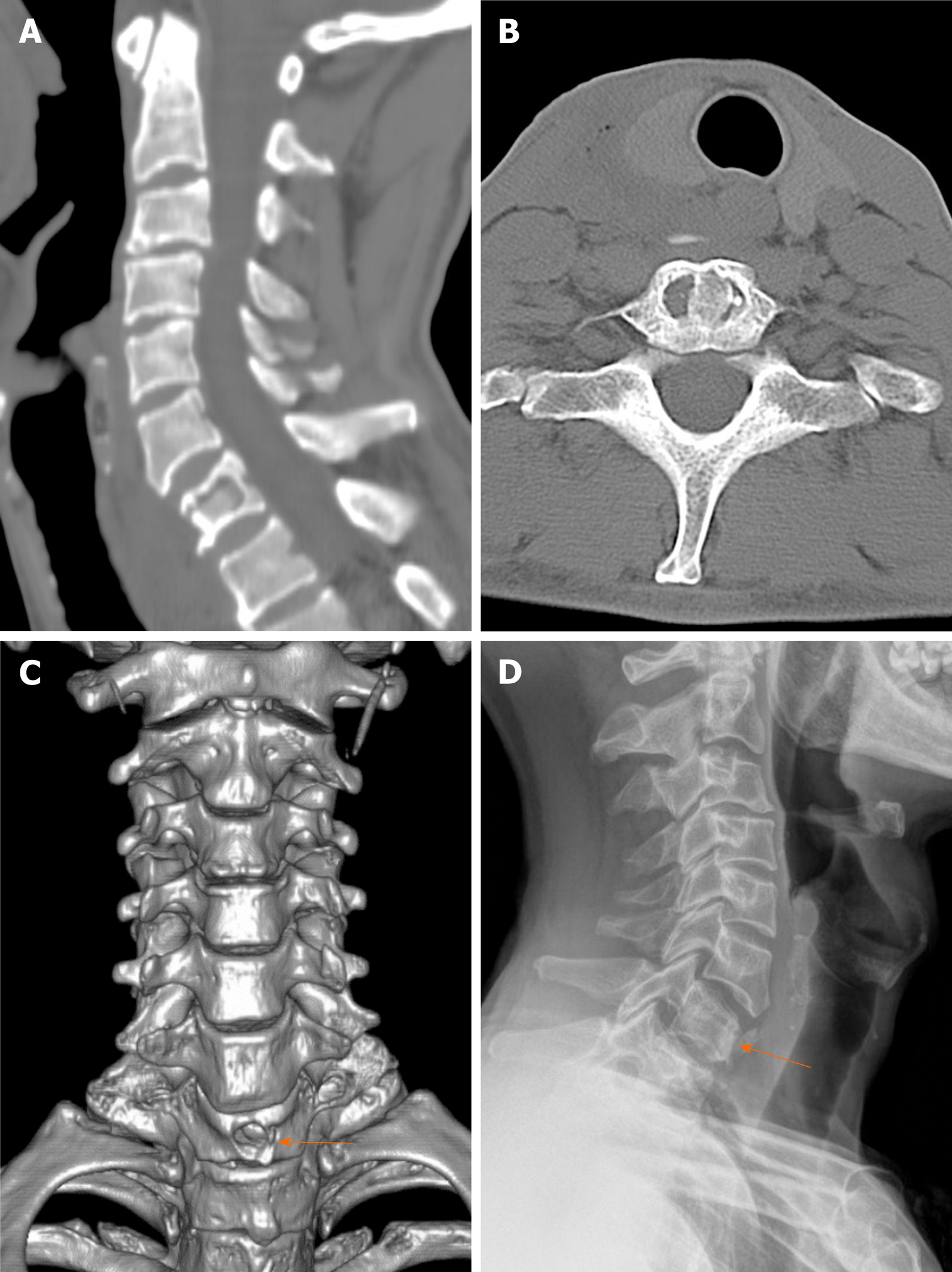
Figure 3 Imaging findings after treatment.
A-C: The axial computed tomography (CT) images (A), sagittal CT images (B) and the Three-dimensional CT reconstruction (C) of the cervical vertebra after the operation showed a good position for bone grafting; D: The last follow-up cervical X-ray showed no radiological recurrence.
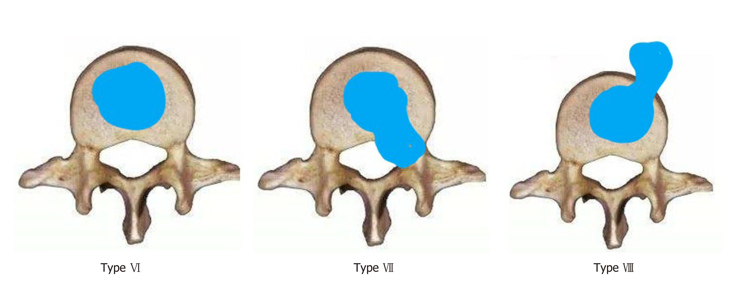
Figure 4 Modified classification of spinal intraosseous schwannomas.
Type VI: Tumors in entirely intravertebral location without intraspinal portion; Type VII: Intraspinal tumor with erosion into vertebral bodies (invasive tumor) and extension into nerve root foramen;Type VIII:Tumors including intraosseous tumors with exophytic component without intraspinal or neural foraminal extension.
- Citation: Xu ZQ, Zhang P, Zhong ZH, Zhou W, Yu HT. Spinal intraosseous schwannoma without spinal canal and neuroforamina involvement: A case report. World J Clin Cases 2020; 8(7): 1271-1277
- URL: https://www.wjgnet.com/2307-8960/full/v8/i7/1271.htm
- DOI: https://dx.doi.org/10.12998/wjcc.v8.i7.1271


