Copyright
©The Author(s) 2020.
World J Clin Cases. Mar 26, 2020; 8(6): 1180-1187
Published online Mar 26, 2020. doi: 10.12998/wjcc.v8.i6.1180
Published online Mar 26, 2020. doi: 10.12998/wjcc.v8.i6.1180
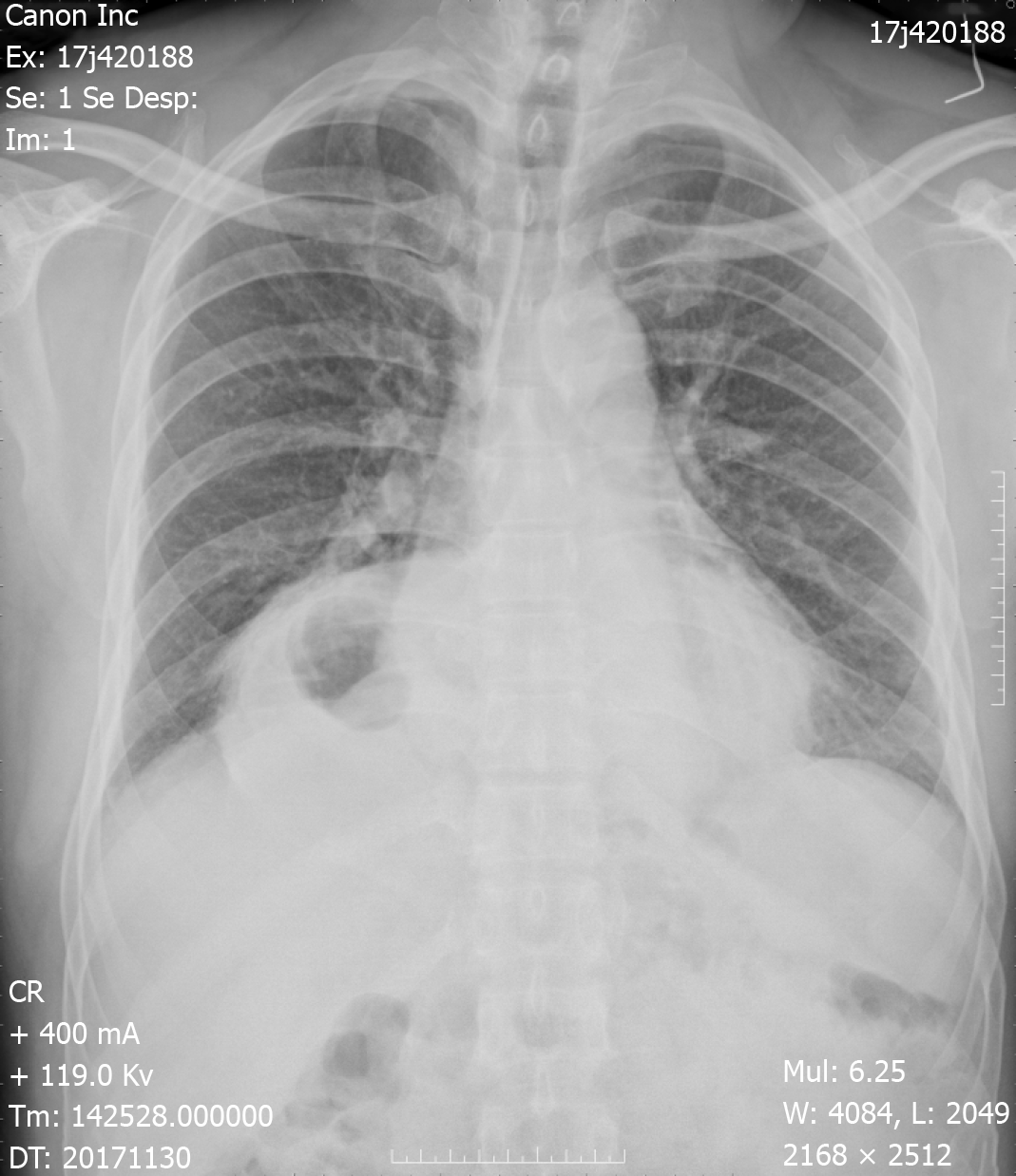
Figure 1 Preoperative chest X-ray.
An intrathoracic gastric bubble with increased bilateral lung marking.
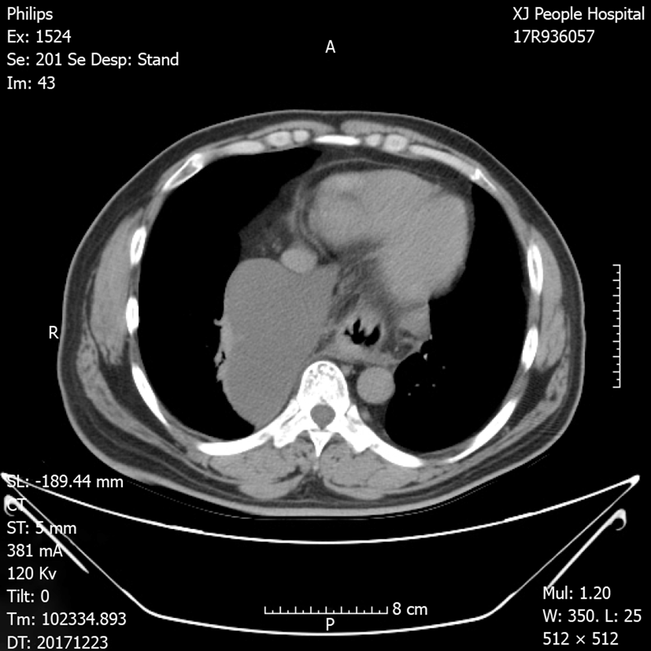
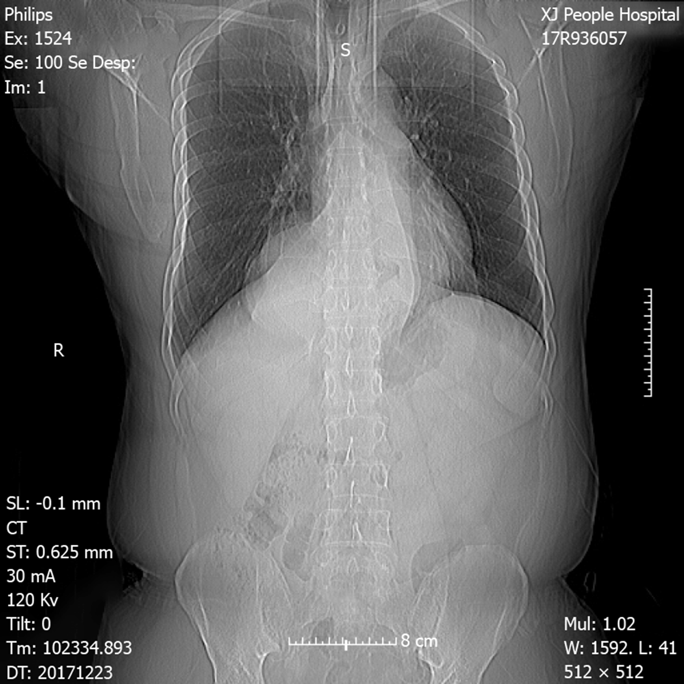
Figure 2 Chest computed tomography with volumetric analysis.
Post-mediastinal location of the whole stomach along with peritoneal fat compressing both bilateral lung and heart.
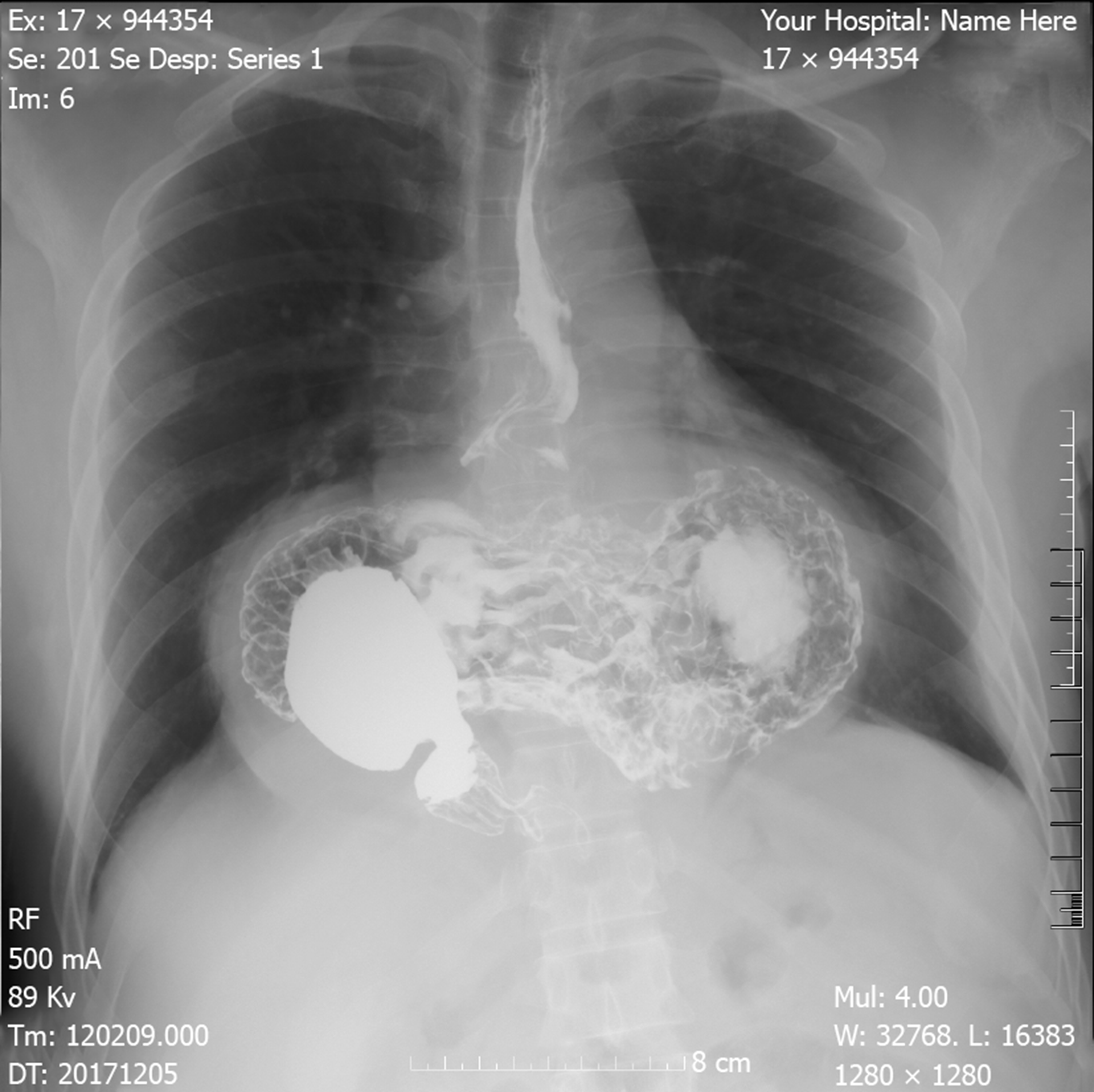
Figure 3 Barium contrast radiography.
Confirming large hiatal hernia, and the configuration of the stomach within the hernia suggested an organoaxial volvulus.
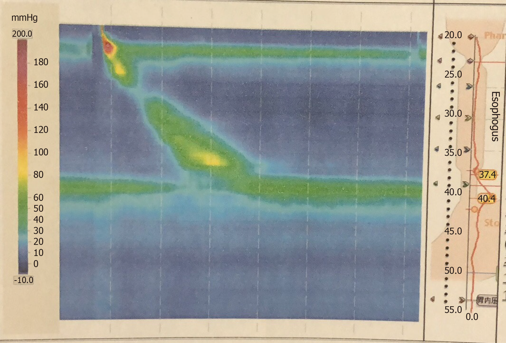
Figure 4 Esophageal high-resolution manometry.
The presence of hiatal hernia with an elevated level of lower esophageal sphincter of 9.6 cm high, pathological acid reflux.
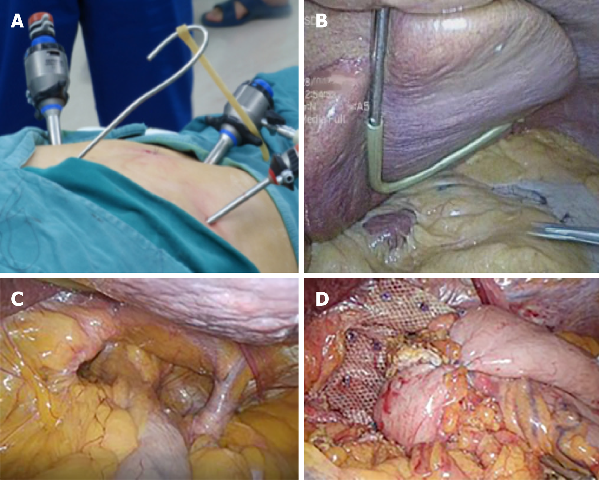
Figure 5 Mesh reinforcement and Nissen fundoplication.
A: The homemade liver retractor. External view of reverse “7” shaped, homemade liver retractor; B: Intra-operative view of the retractor; C: Repositioning of intra-abdominal esophagus. Three centimeters of tension-free esophagus was repositioned intra-abdominally; D: Three hundred sixty degree Nissen fundoplication and mesh reinforcement.
Figure 6 Postoperative chest x-ray at 1-mo follow up.
There were no distinct abnormalities observed.
- Citation: Yasheng D, Wulamu W, Li YL, Tuhongjiang A, Abudureyimu K. Laparoscopic repair of complete intrathoracic stomach with iron deficiency anemia: A case report. World J Clin Cases 2020; 8(6): 1180-1187
- URL: https://www.wjgnet.com/2307-8960/full/v8/i6/1180.htm
- DOI: https://dx.doi.org/10.12998/wjcc.v8.i6.1180


