Copyright
©The Author(s) 2020.
World J Clin Cases. Mar 26, 2020; 8(6): 1150-1157
Published online Mar 26, 2020. doi: 10.12998/wjcc.v8.i6.1150
Published online Mar 26, 2020. doi: 10.12998/wjcc.v8.i6.1150
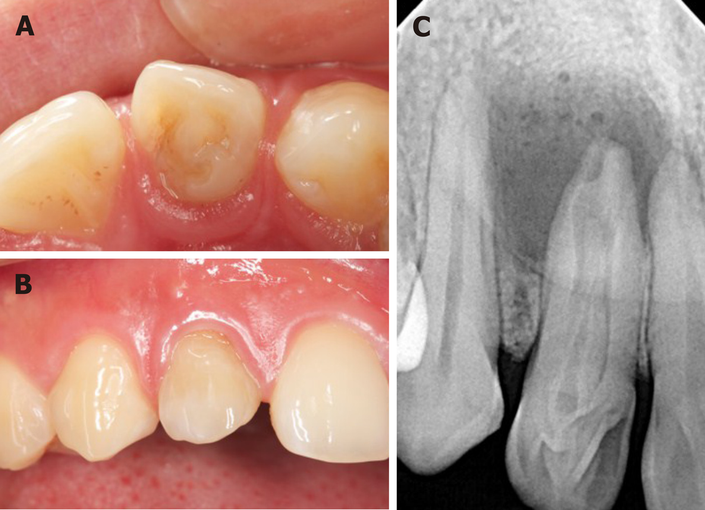
Figure 1 The intraoral examination before treatment.
A: The lingual view: the tooth has unusual morphologic features; B: The labial view: the upper right lateral tooth is slightly discolored; C: A preoperative radiograph of the tooth showed a type III dens invaginatus and an associated large apical radiolucency.
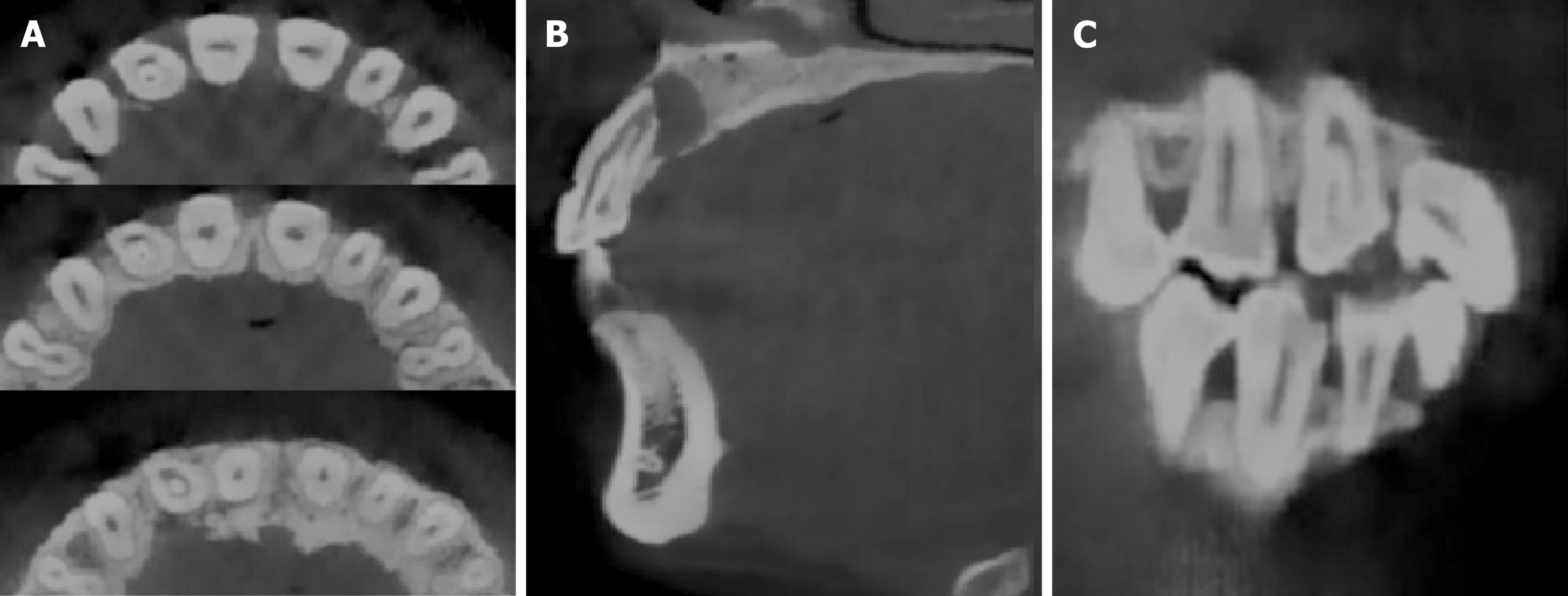
Figure 2 Cone-beam computed tomography images of dens invaginatus.
A: The axial image; B: The sagittal image; C: The coronal sections image. A type III dens invaginatus tooth with apical periodontitis based on Oehlers classification can easily be seen in the images.
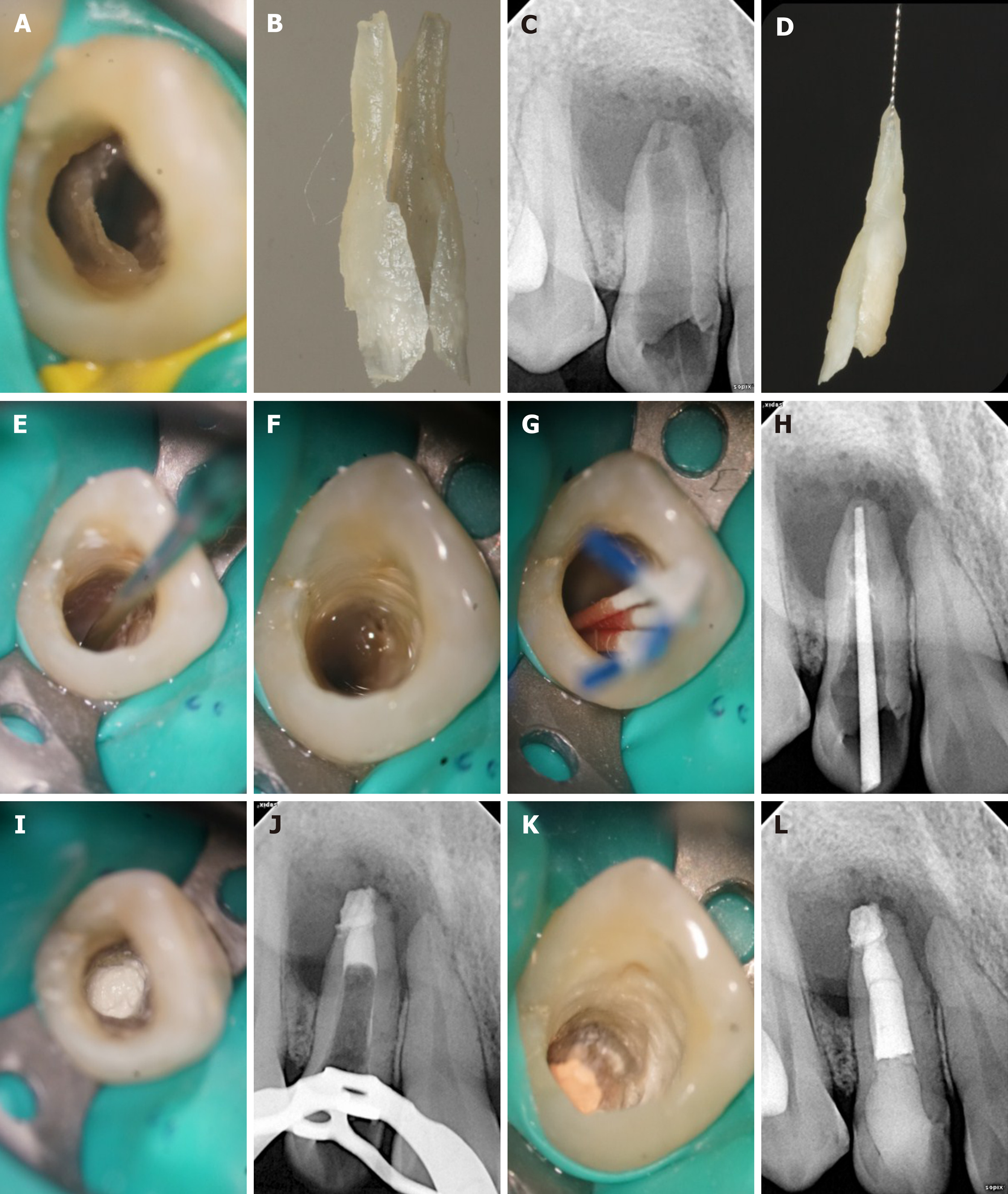
Figure 3 Non-surgical treatment process.
A: Invaginated tooth seen under the dental operation microscope; B: Partially invaginated tooth taken out for the first step; C: Intraoperative film confirmed that the invaginated tooth was completely removed; D: In vitro photo of a complete invaginated tooth; E: The canal was prepared with ultrasonic tip; F: The canal was irrigated with 1% sodium hypochlorite and saline; G: The canal was dried with absorbent paper points; H: Working length was confirmed with the X-ray dental pictures; I: Mineral trioxide aggregate was seen under the dental operation microscope; J: A radiographic image showed the apical third root was obturated with 4 mm mineral trioxide aggregate; K: The remaining pulp space was obturated with thermoplastic gutta-percha, and the access cavity was sealed with a composite resin; L: Radiograph taken immediately after the treatment.
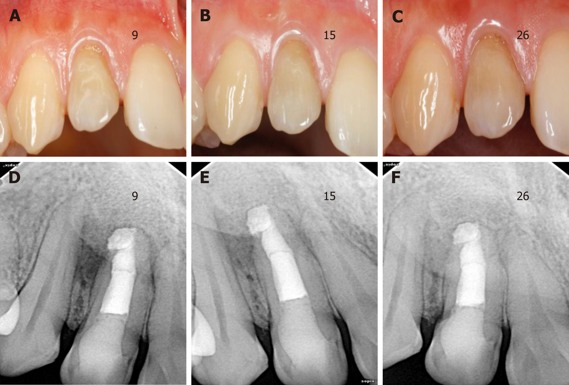
Figure 4 Follow-up images at different periods.
A-C: The labial view of the invaginated tooth during 9-, 15- and 26-mo follow-up examinations; D-F: Periapical radiographs taken at the 9-, 15- and 26-mo follow-up examinations. The apical radiolucency was significantly reduced.
- Citation: Liu J, Zhang YR, Zhang FY, Zhang GD, Xu H. Microscopic removal of type III dens invaginatus and preparation of apical barrier with mineral trioxide aggregate in a maxillary lateral incisor: A case report and review of literature. World J Clin Cases 2020; 8(6): 1150-1157
- URL: https://www.wjgnet.com/2307-8960/full/v8/i6/1150.htm
- DOI: https://dx.doi.org/10.12998/wjcc.v8.i6.1150


