Copyright
©The Author(s) 2020.
World J Clin Cases. Mar 26, 2020; 8(6): 1129-1136
Published online Mar 26, 2020. doi: 10.12998/wjcc.v8.i6.1129
Published online Mar 26, 2020. doi: 10.12998/wjcc.v8.i6.1129
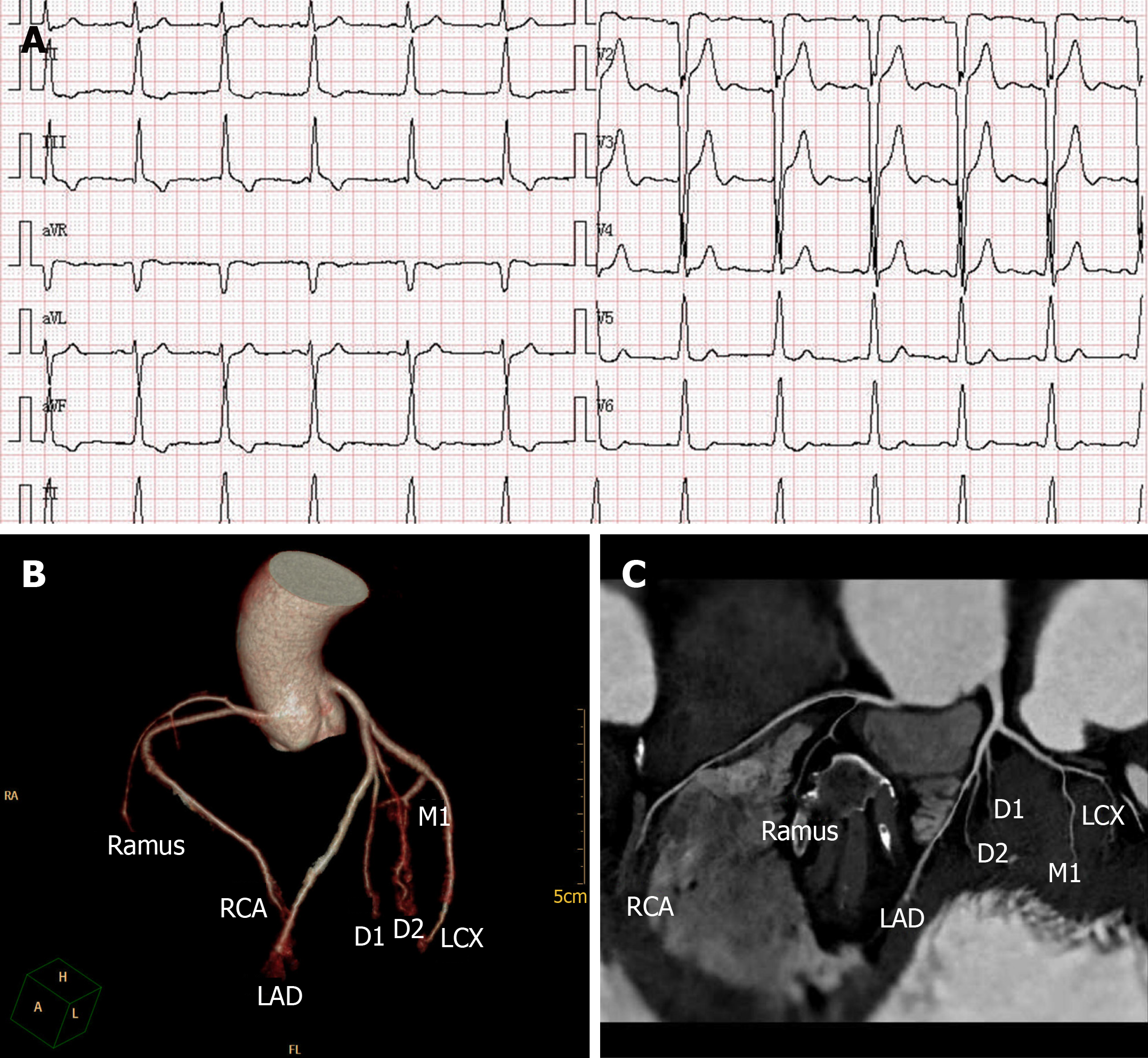
Figure 1 Electrocardiogram and coronary computed tomography angiography.
A: Twelve-lead electrocardiogram revealed a sinus rhythm with QS waves, an intraventricular conduction block (QRS duration, 118 ms), and a change of the ST-T wave in 2017. B and C: Coronary computed tomography angiography showed no abnormality in our patient. LAD: Left anterior descending; LCX: Left circumflex coronary artery; RCA: Right coronary artery; D1, D2: Diagonal branches; M1: Marginal branches.
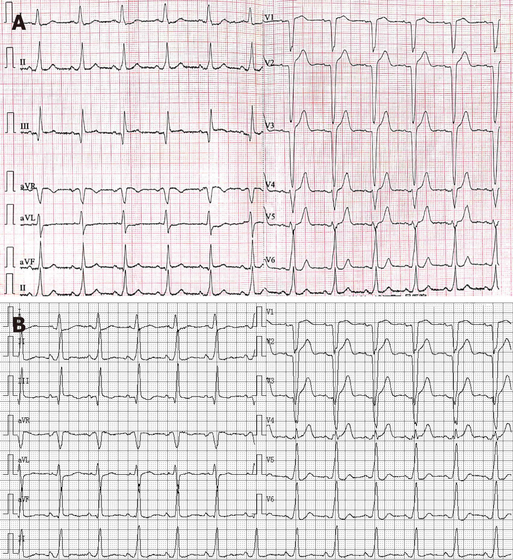
Figure 2 Electrocardiogram.
A and B: Twelve-lead electrocardiograms revealed monomorphic wide QRS complex ventricular tachycardia at an emergency room of a local hospital in June and July 2019.
Figure 3 Electrocardiogram.
A: Twelve-lead electrocardiogram revealed a sinus rhythm and left bundle branch block (LBBB) (QRS duration, 143 ms) at an emergency room of a local hospital; B: Twelve-lead electrocardiogram revealed a sinus rhythm and LBBB (QRS duration, 126 ms) at rest.
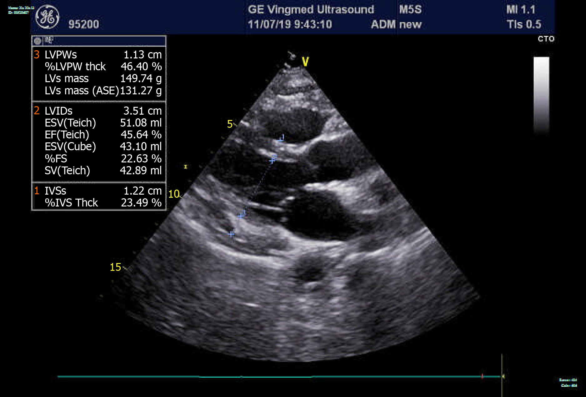
Figure 4 Echocardiogram.
Echocardiogram pre-operation showed a normal left ventricular ejection fraction of 58% and normal cardiac chamber size with no structural abnormality of both ventricles. LVPWs: Left ventricular posterior wall systolic thickness; LVPW: Left ventricular posterior wall; LVs mass: Left ventricular end-systolic mass; LVIDs: Left ventricle internal diameter at end-systole; ESV: End-systolic volume; EF: Ejection fraction; FS: Fractional shortening; SV: Systolic volume; IVSs: Interventricular septum end-systolic thickness; IVS: Interventricular septum.
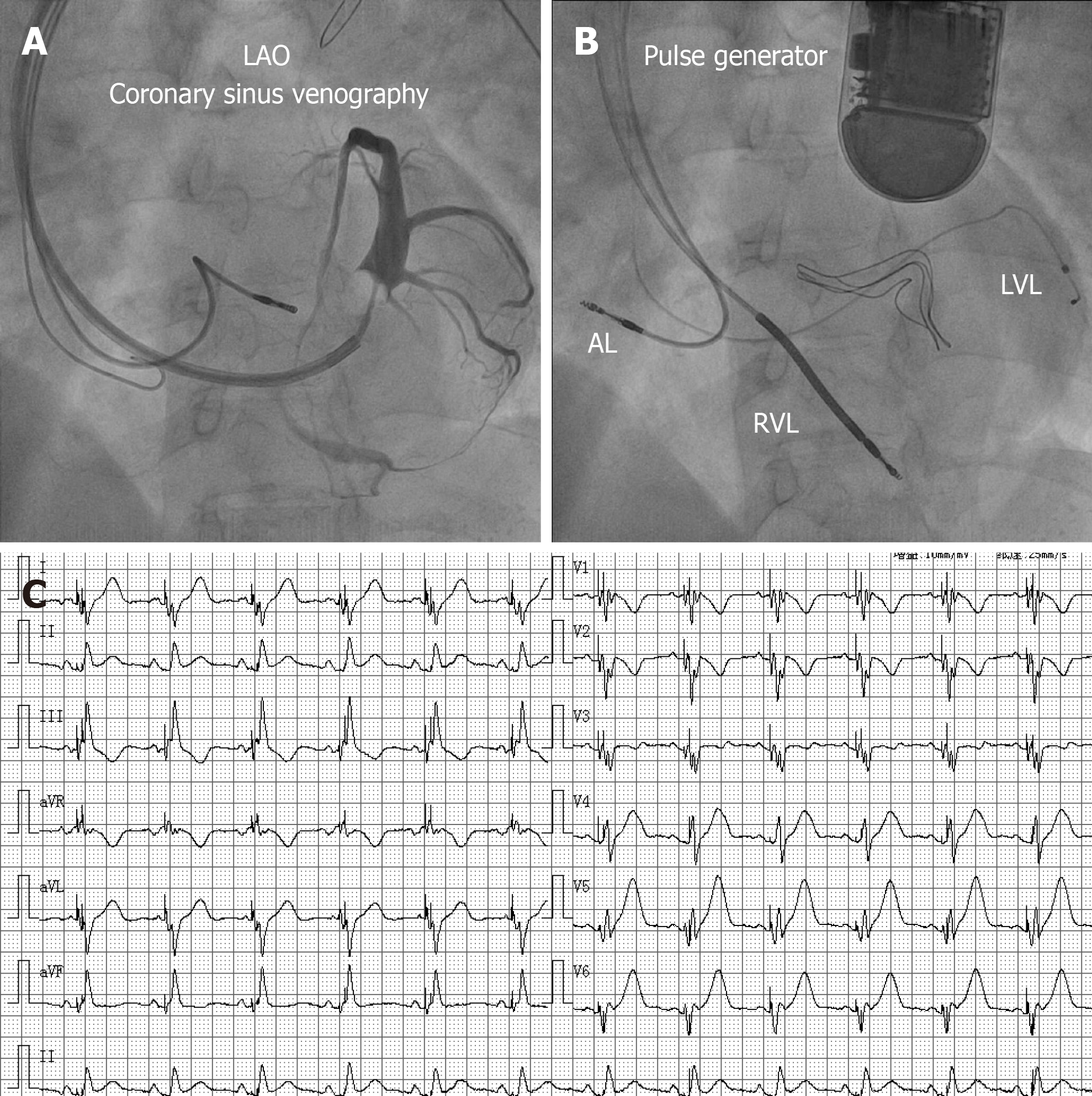
Figure 5 Coronary sinus venography and electrocardiogram.
A: Coronary sinus venography performed from a left anterior oblique 30°angle, and presentation of the coronary vein; B: Anteroposterior view of the position of the final leads; C: Twelve-lead electrocardiograms revealed biventricular pacing and a QRS duration of 104 ms after successful implantation of a cardiac resynchronization therapy-defibrillator. AL: Atrial lead; RVL: Right ventricular lead; LVL: Left ventricular lead. LAO: Left anterior oblique.
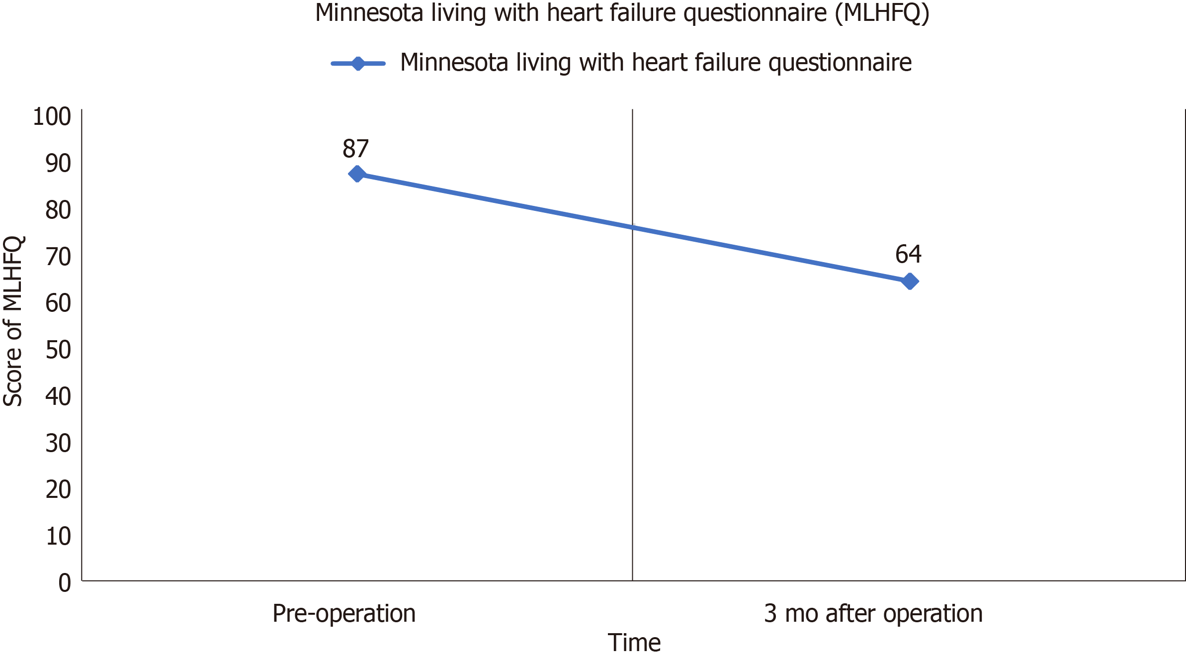
Figure 6 Minnesota living with heart failure questionnaire.
Minnesota living with heart failure questionnaire score decreased from 87 to 64 at the 3-mo follow-up.
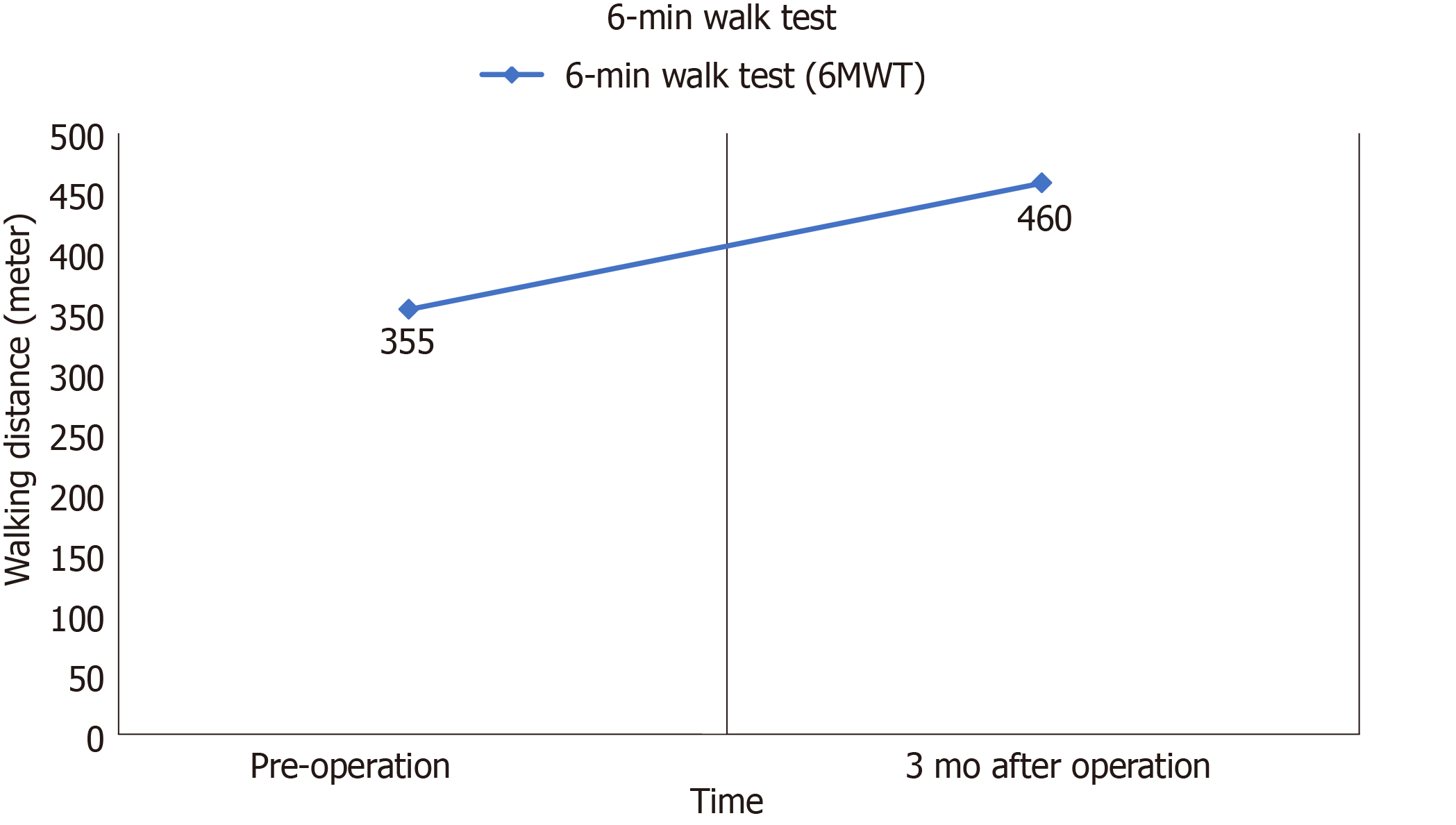
Figure 7 6-min walk test.
Walking distance of 6-min walk test was elevated from 355 to 460 meters at the 3-mo follow-up.
- Citation: Chen YY, Yan H, Zhu JH. Successful treatment of systemic sclerosis complicated by ventricular tachycardia with a cardiac resynchronization therapy-defibrillator: A case report. World J Clin Cases 2020; 8(6): 1129-1136
- URL: https://www.wjgnet.com/2307-8960/full/v8/i6/1129.htm
- DOI: https://dx.doi.org/10.12998/wjcc.v8.i6.1129


