Copyright
©The Author(s) 2020.
World J Clin Cases. Feb 6, 2020; 8(3): 535-539
Published online Feb 6, 2020. doi: 10.12998/wjcc.v8.i3.535
Published online Feb 6, 2020. doi: 10.12998/wjcc.v8.i3.535
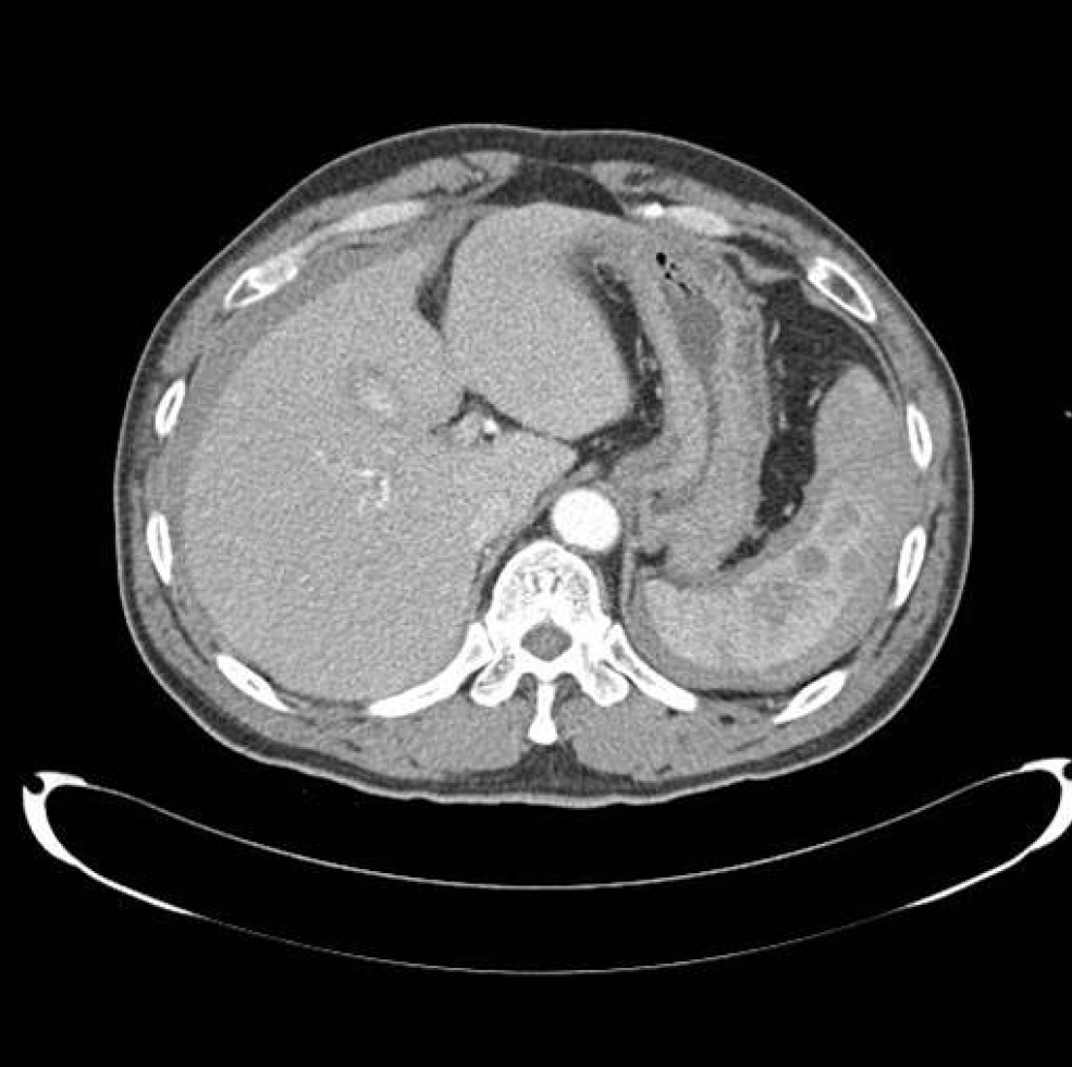
Figure 1 Computed tomography shows multiple hemorrhagic cysts in the spleen and a moderate amount of hemoperitoneum.
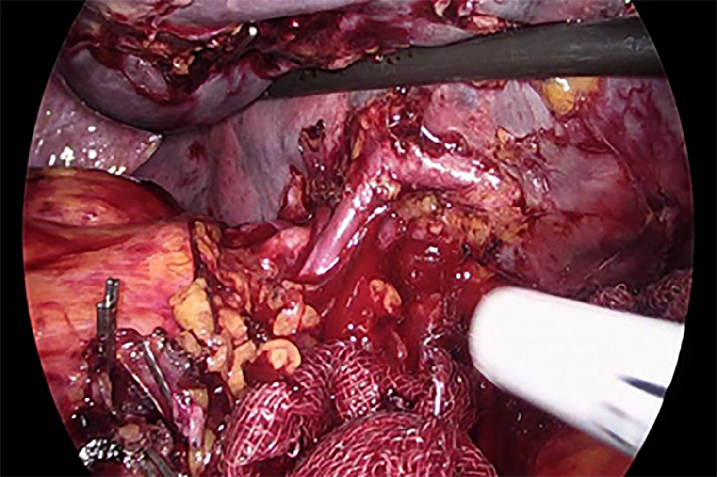
Figure 2 Tortuous and overdeveloped vessels in surgical field.
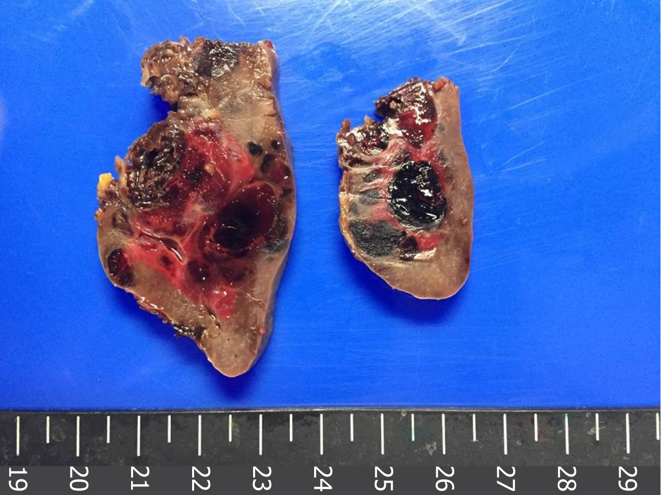
Figure 3 Gross image of the specimen.
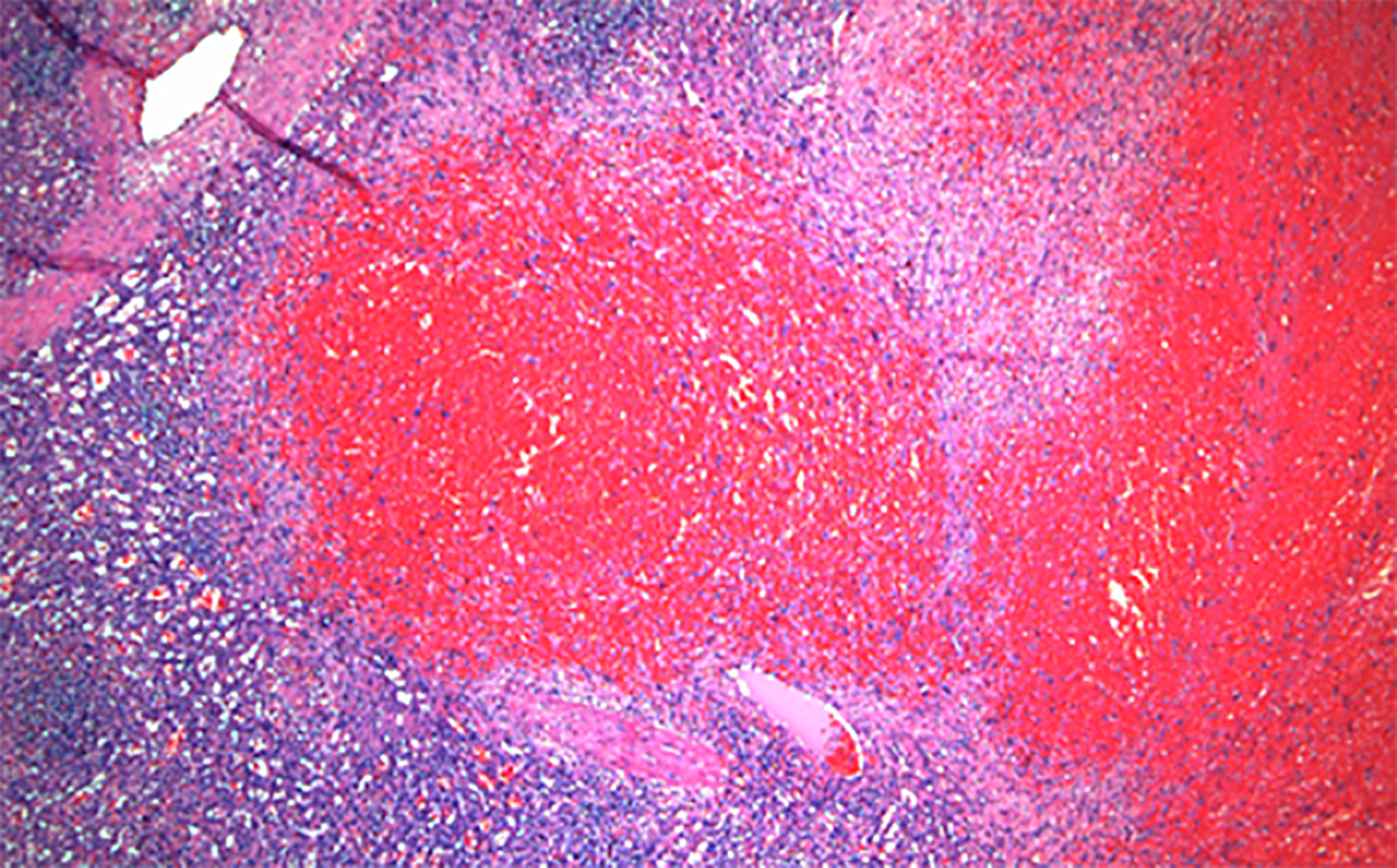
Figure 4 Microscopic image of the specimen.
- Citation: Rhu J, Cho J. Ruptured splenic peliosis in a patient with no comorbidity: A case report. World J Clin Cases 2020; 8(3): 535-539
- URL: https://www.wjgnet.com/2307-8960/full/v8/i3/535.htm
- DOI: https://dx.doi.org/10.12998/wjcc.v8.i3.535


