Copyright
©The Author(s) 2020.
World J Clin Cases. Dec 26, 2020; 8(24): 6537-6545
Published online Dec 26, 2020. doi: 10.12998/wjcc.v8.i24.6537
Published online Dec 26, 2020. doi: 10.12998/wjcc.v8.i24.6537
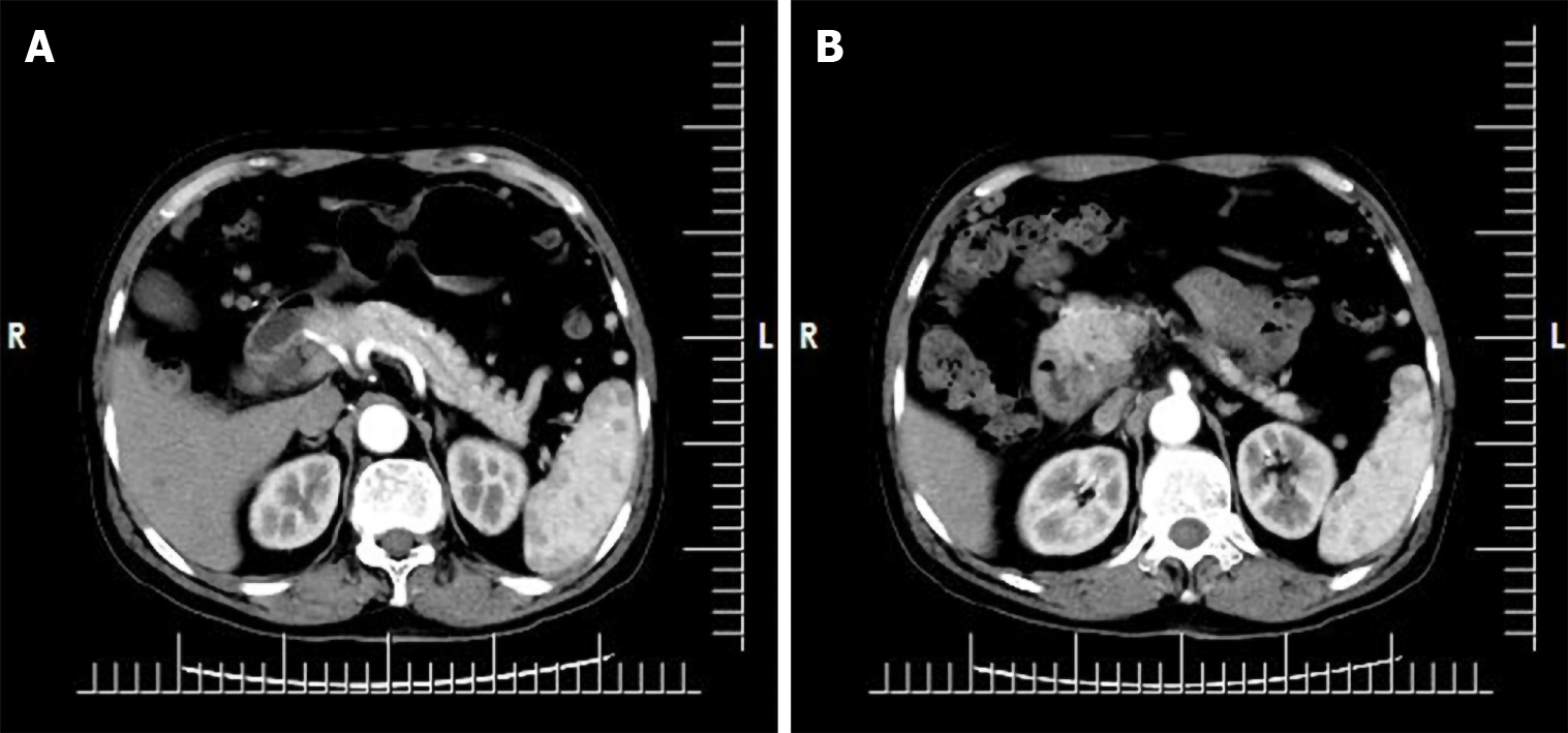
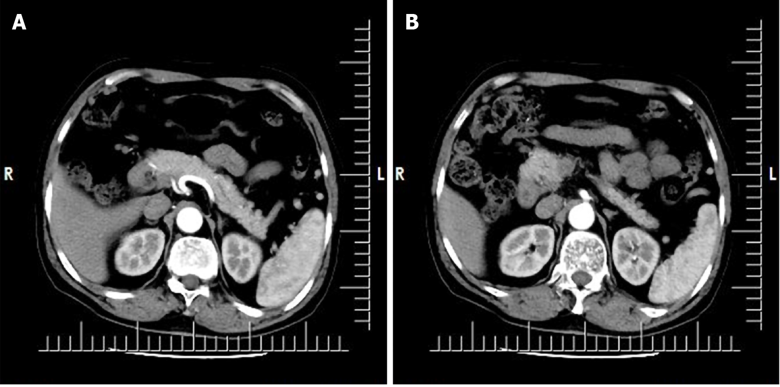
Figure 1 Upper abdominal enhanced computed tomography at Beijing Dong Fang Hospital.
A: The wall of the descending part of the duodenum showed local thickening, with an iso-low density shadow and blurred edge; B: Enhanced scan showed obvious non-uniform enhancement. The impression diagnosis was space-occupying lesion in the descending part of the duodenum, and malignancy was considered.
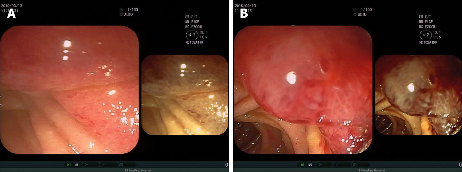
Figure 2 Electronic gastroscope findings.
Neoplasms were seen on the medial wall of the descending part of the duodenum, which showed obvious hyperemia and irregular shape. The nature of the findings was to be determined. A: The junction of the proliferative lesion and duodenal wall; B: The surface shape of the proliferative lesion was irregular, and hyperemia was obvious.
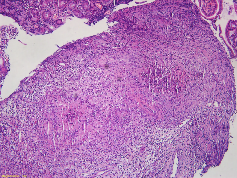
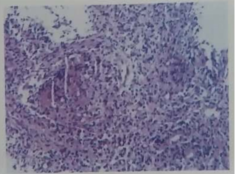
Figure 3 Duodenal mucosa biopsy stained with hematoxylin-eosin.
Magnification is 10 ×.
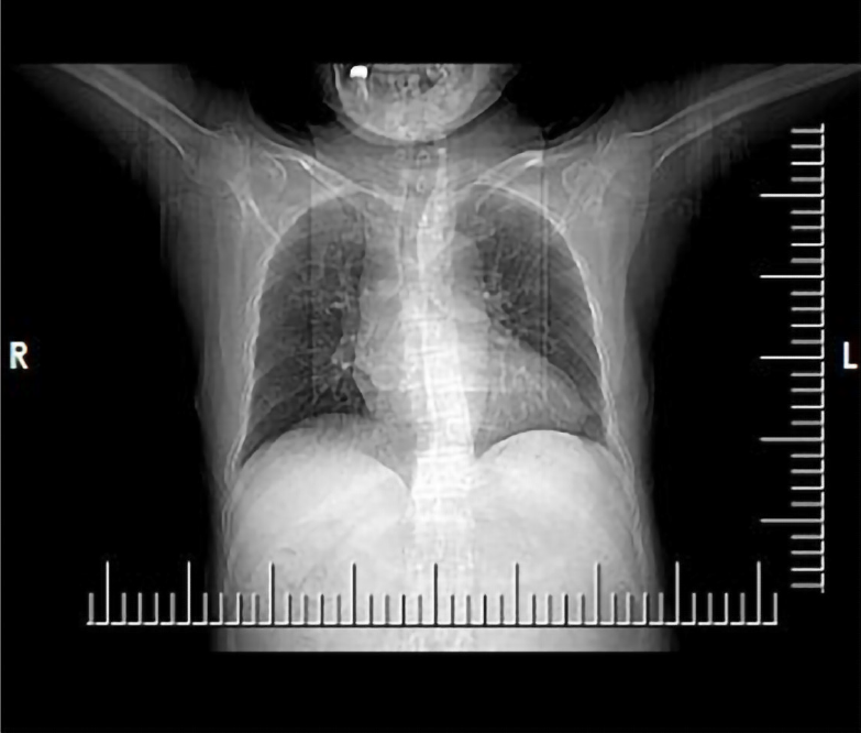
Figure 4 Chest computed tomography plain scan showing bronchiectasis with infection in both lungs.
No obvious characteristics of pulmonary tuberculosis were apparent.
Figure 5 Finding presented in the report on pathological consultation in the Beijing Chest Hospital.
The descending duodenum tissue showed chronic granulomatous inflammation, with slight necrosis and suppuration. The lesion was deemed consistent with a diagnosis of tuberculosis.
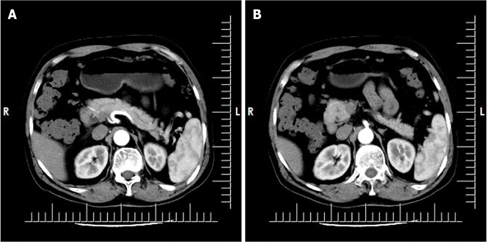
Figure 6 Upper abdominal computed tomography after 1 mo of anti-tuberculosis treatment.
There was no significant change in the location of the duodenum. A and B: Representative images at different levels.
Figure 7 Upper abdominal computed tomography after 6 mo of anti-tuberculosis treatment.
There was no significant change in the location of the duodenum. A and B: Representative images at different levels.
- Citation: Zhang Y, Shi XJ, Zhang XC, Zhao XJ, Li JX, Wang LH, Xie CE, Liu YY, Wang YL. Primary duodenal tuberculosis misdiagnosed as tumor by imaging examination: A case report. World J Clin Cases 2020; 8(24): 6537-6545
- URL: https://www.wjgnet.com/2307-8960/full/v8/i24/6537.htm
- DOI: https://dx.doi.org/10.12998/wjcc.v8.i24.6537


