Copyright
©The Author(s) 2020.
World J Clin Cases. Dec 26, 2020; 8(24): 6529-6536
Published online Dec 26, 2020. doi: 10.12998/wjcc.v8.i24.6529
Published online Dec 26, 2020. doi: 10.12998/wjcc.v8.i24.6529
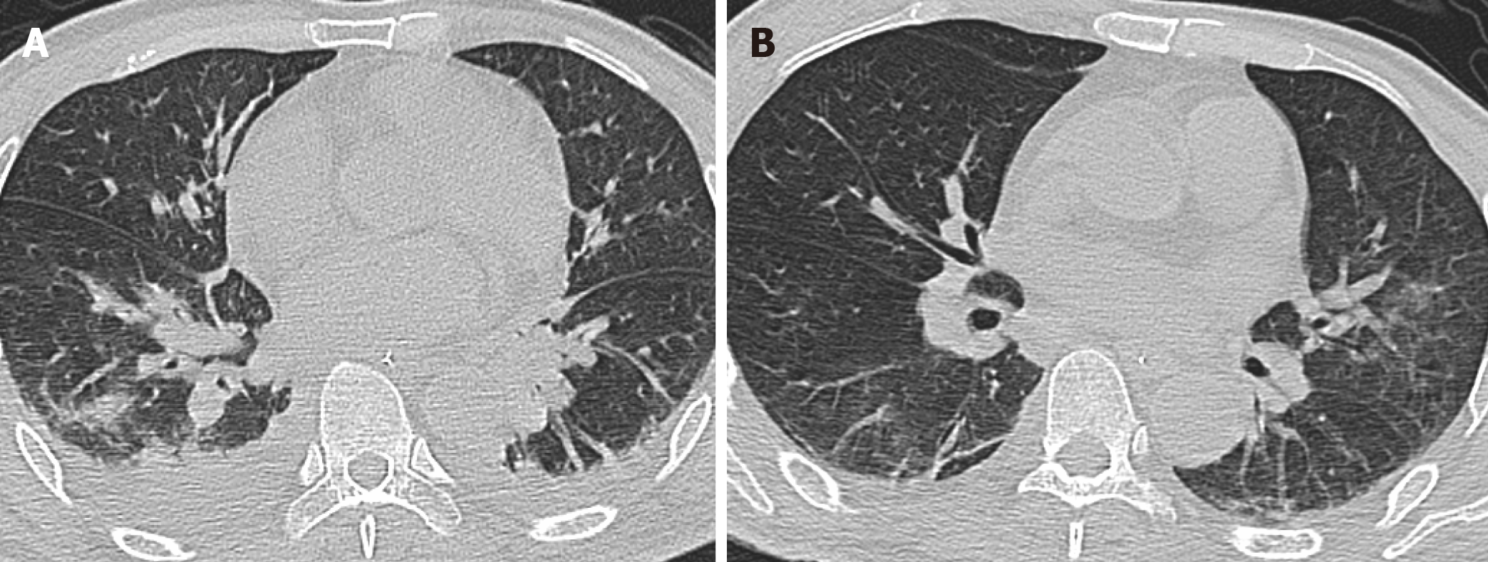
Figure 1 Findings of chest computed tomography.
A: Three weeks before the patient was transferred to our hospital, chest computed tomography showed pulmonary consolidation and pleural effusion; B: Upon admission to our hospital, chest computed tomography showed that pulmonary infection had alleviated, but some inflammatory infiltrations were present.
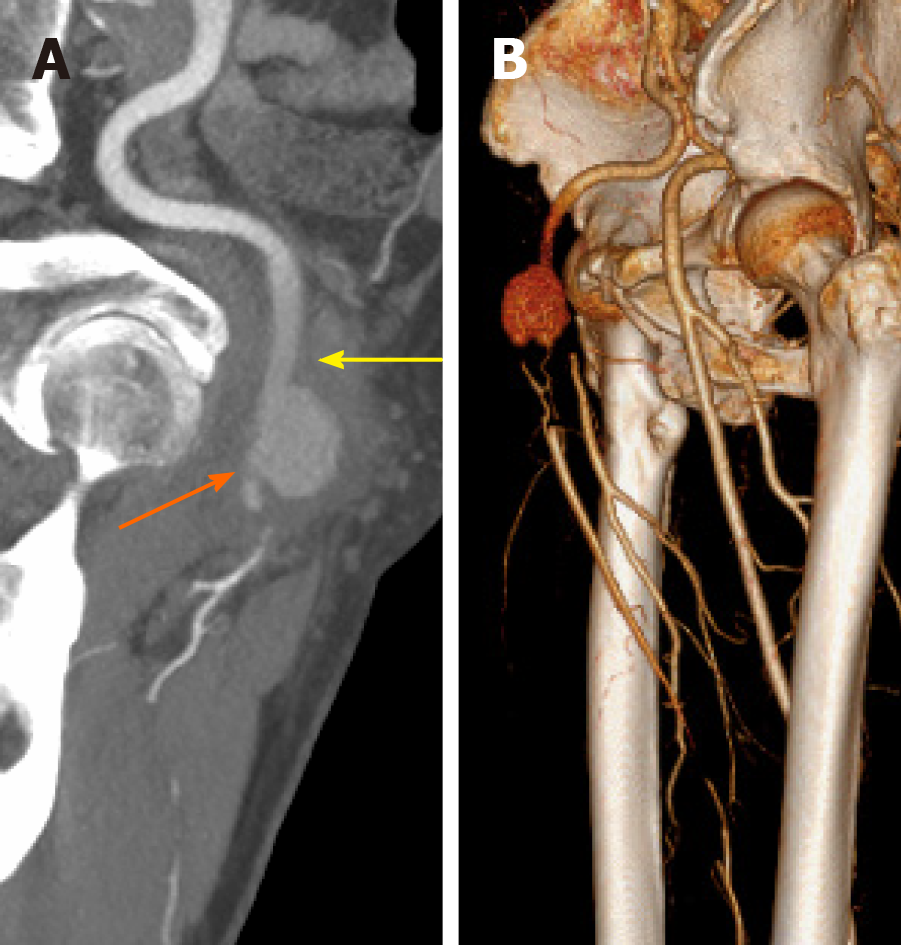
Figure 2 Preoperative computed tomography angiography.
A: A pseudoaneurysm of the common femoral artery compressed its distal outflow tract (orange arrow); B: Occlusion of the superficial femoral artery and deep femoral artery.
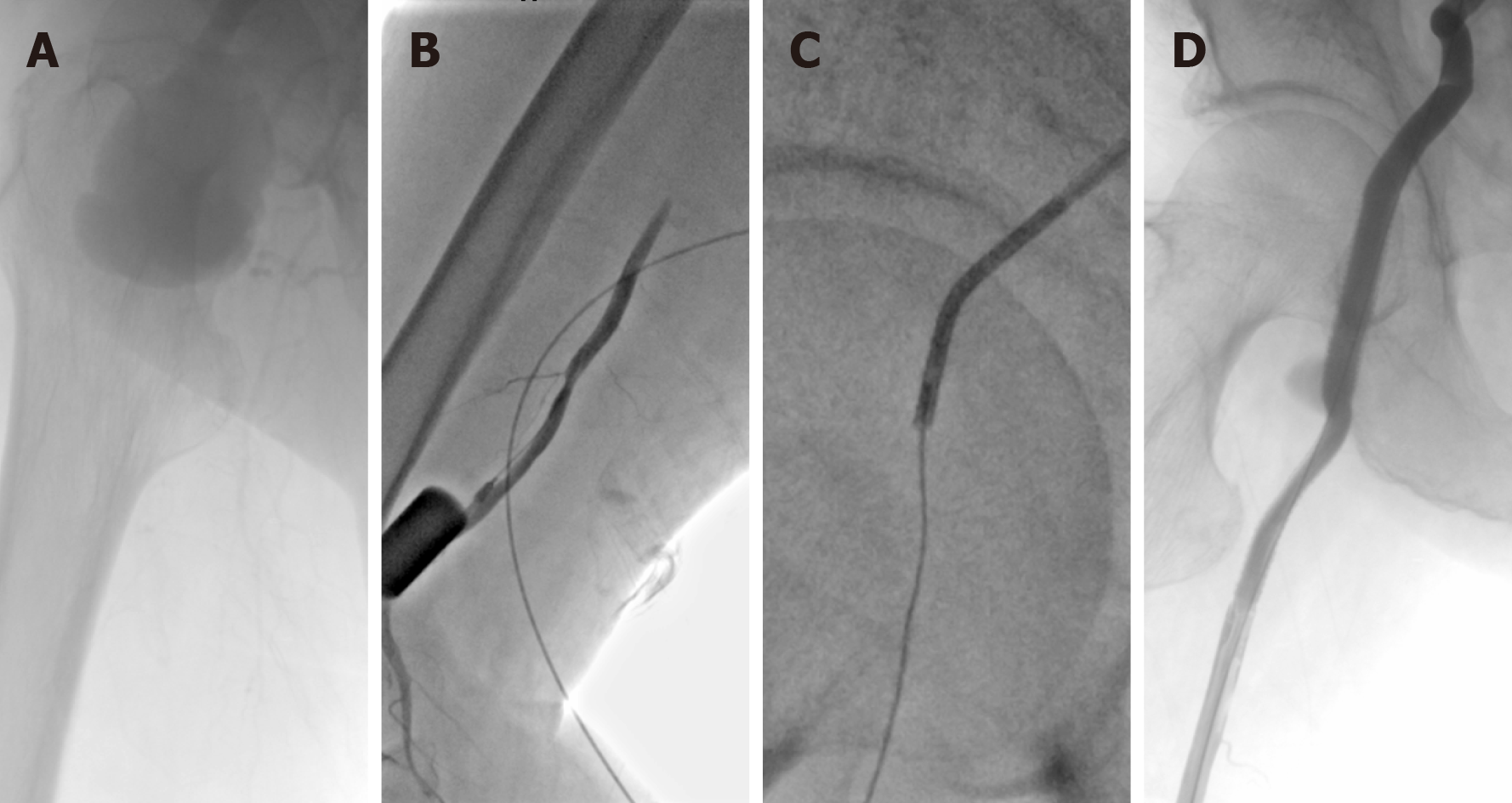
Figure 3 Intraoperative digital subtraction angiography.
A: Guidewires cannot pass through the occlusion lesion; B: Retrograde access was achieved at the superficial femoral artery; C: Pathway was established via the “SAFARI” technique; D: After stent–grafts were deployed, the stent–grafts and distal outflow tract were patent without any endoleaks.
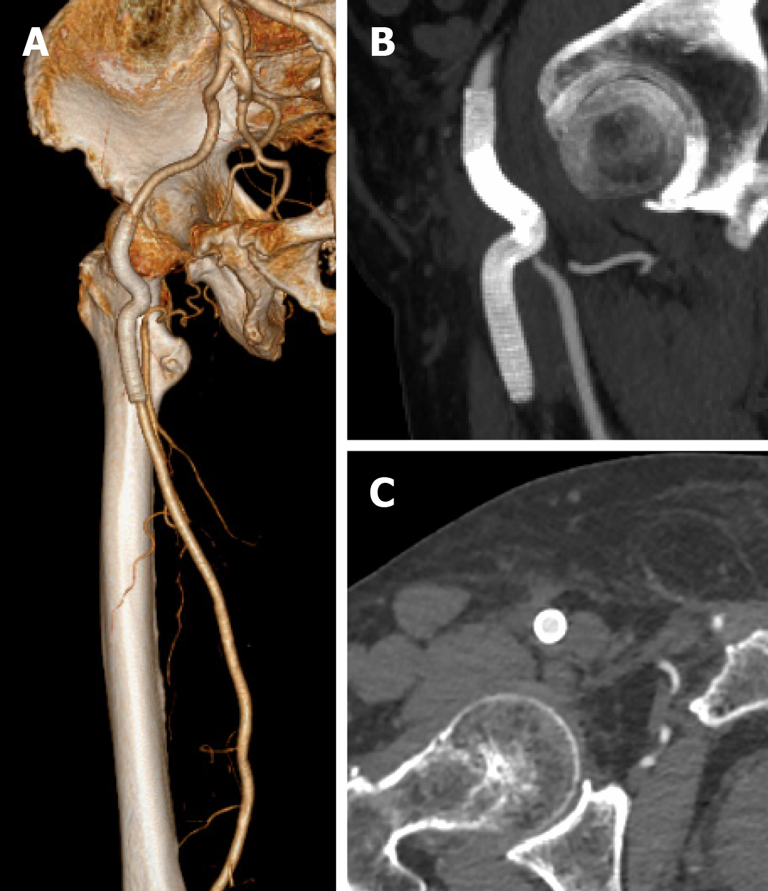
Figure 4 Computed tomography angiography at the 1-year follow-up.
A: Stent–grafts were patent without any endoleaks; B and C: The previous surrounding hematoma was completely absorbed.
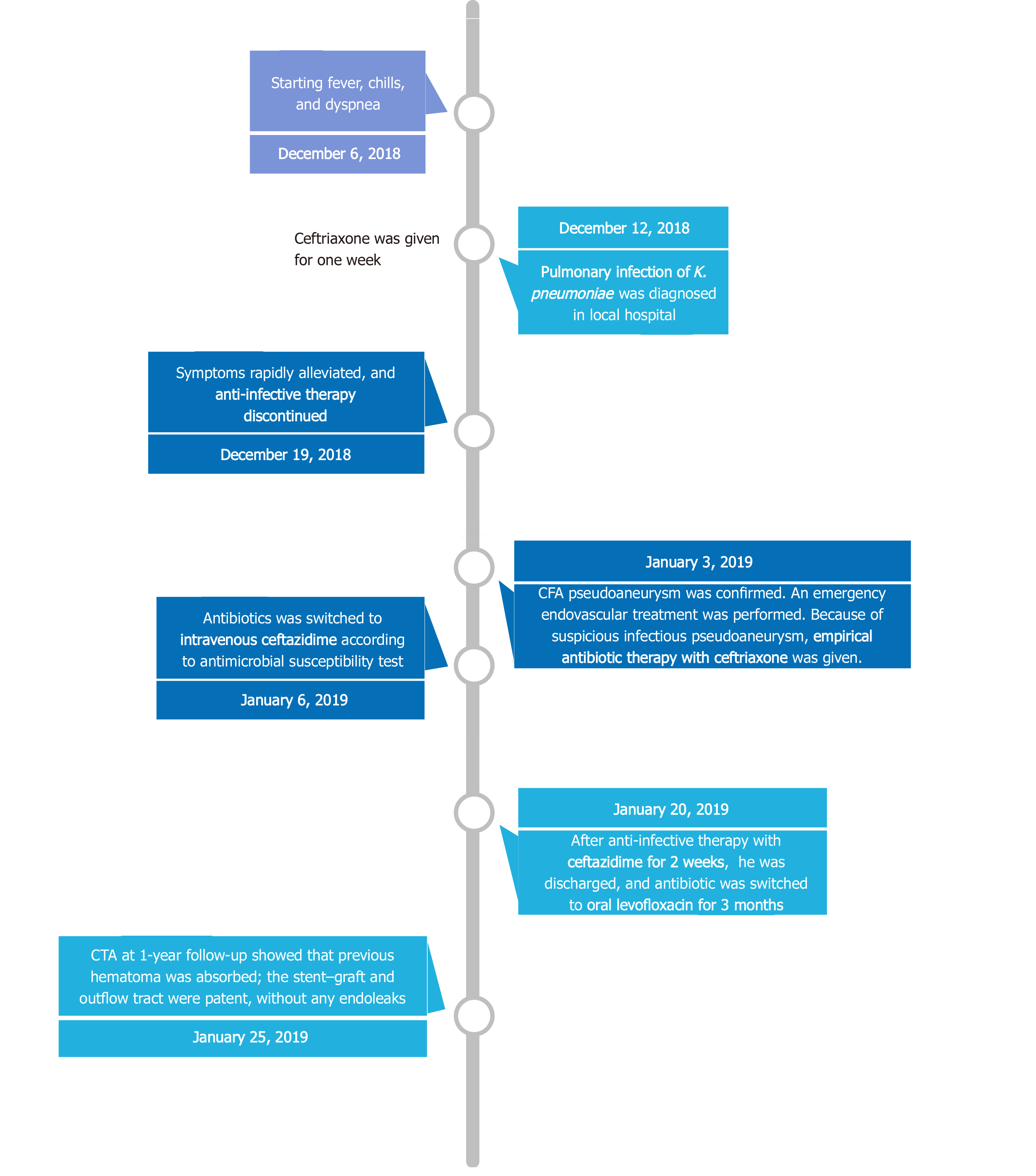
Figure 5 Timeline of the treatment.
CFA: Common femoral artery; CTA: Computed tomography angiography.
- Citation: Wang TH, Zhao JC, Huang B, Wang JR, Yuan D. Successful endovascular treatment with long-term antibiotic therapy for infectious pseudoaneurysm due to Klebsiella pneumoniae: A case report. World J Clin Cases 2020; 8(24): 6529-6536
- URL: https://www.wjgnet.com/2307-8960/full/v8/i24/6529.htm
- DOI: https://dx.doi.org/10.12998/wjcc.v8.i24.6529


