Copyright
©The Author(s) 2020.
World J Clin Cases. Dec 26, 2020; 8(24): 6504-6510
Published online Dec 26, 2020. doi: 10.12998/wjcc.v8.i24.6504
Published online Dec 26, 2020. doi: 10.12998/wjcc.v8.i24.6504
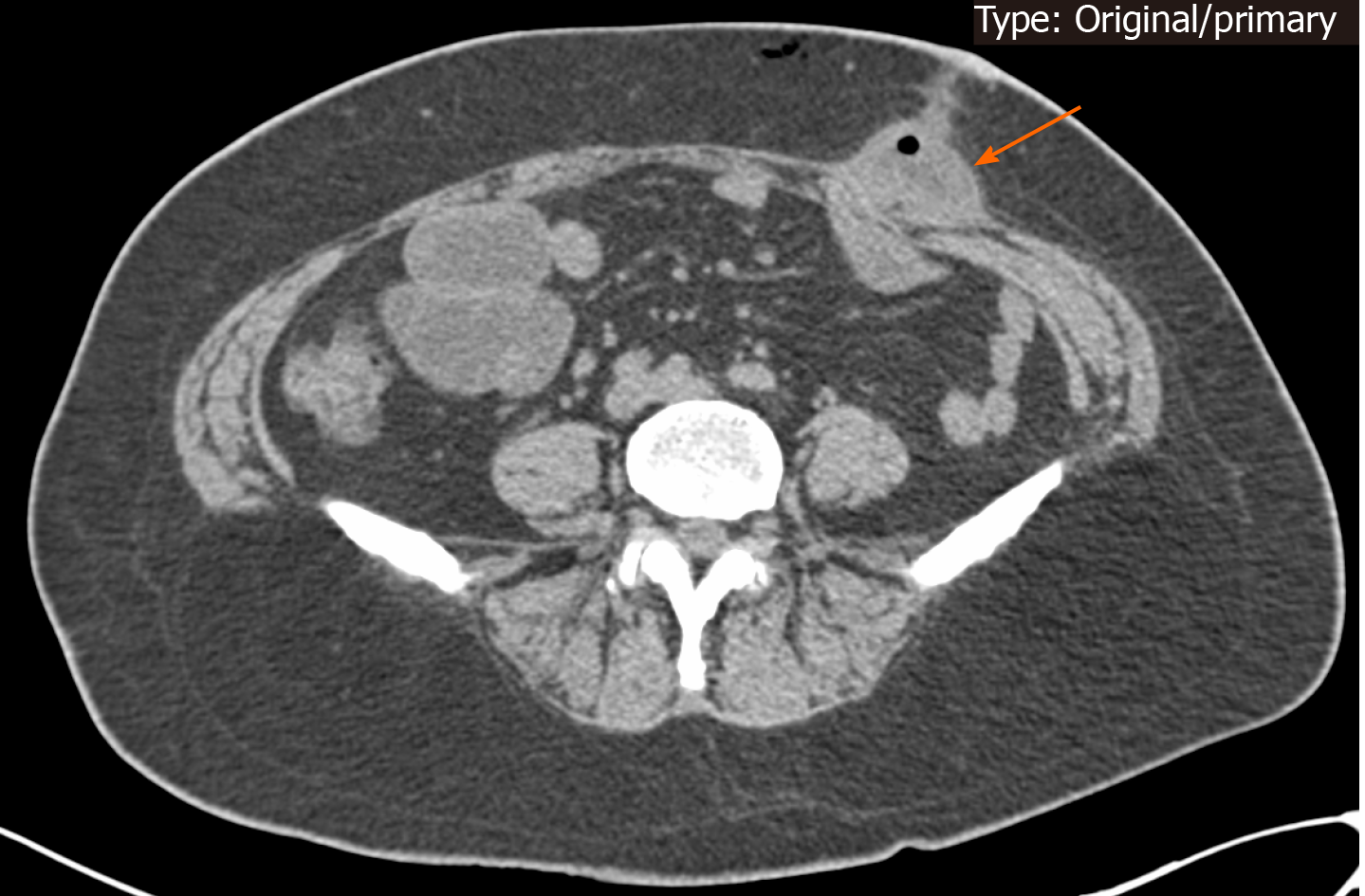
Figure 1 Postoperative day 8 coronal computed tomography scan showing small bowel herniated through the drainage port site.
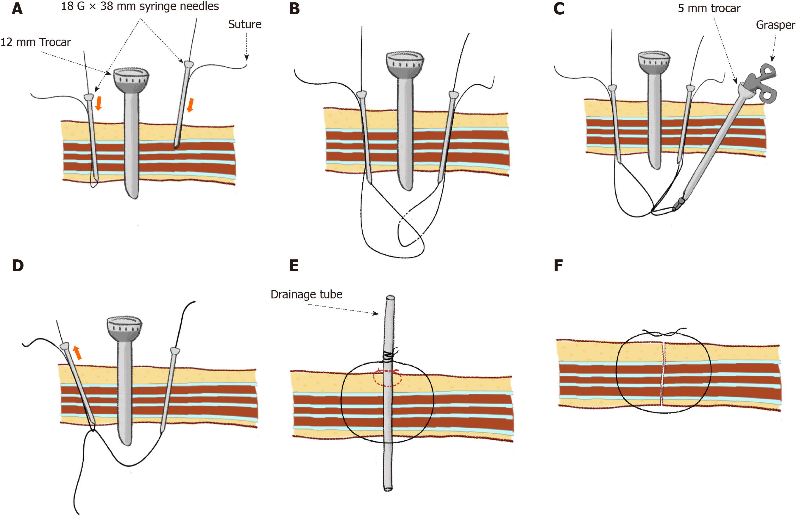
Figure 2 A simple and practical method for closing the trocar incision at the drain-site.
A: Let one end of the suture pass through a syringe needle (18 G × 38 mm), and let the needle penetrate through the whole abdominal wall at one edge of the incision under direct vision. Perform the same way on the other side of the incision; B: Let the right loop trap in the left loop with the help of a grasper; C: Continuously grasp the right loop with a grasper; D: Then, hold one end of the right suture and withdraw the left loop outward. Release the grasper when the right suture is pulled into the left needle and allow the other end of right suture pass to through the position of the left syringe needle. Finally, the right suture crosses all layers of the abdominal wall at the incision site; E: Secure the drainage tube to the skin with a simple skin suture (dotted red line). Then, the right suture is wrapped around the drainage tube during the placement of the drainage tube; F: Finally, the right suture is tied after the drainage tube is removed (also known as delayed closure technique).
- Citation: Gao X, Chen Q, Wang C, Yu YY, Yang L, Zhou ZG. Rare case of drain-site hernia after laparoscopic surgery and a novel strategy of prevention: A case report. World J Clin Cases 2020; 8(24): 6504-6510
- URL: https://www.wjgnet.com/2307-8960/full/v8/i24/6504.htm
- DOI: https://dx.doi.org/10.12998/wjcc.v8.i24.6504


