Copyright
©The Author(s) 2020.
World J Clin Cases. Dec 26, 2020; 8(24): 6473-6479
Published online Dec 26, 2020. doi: 10.12998/wjcc.v8.i24.6473
Published online Dec 26, 2020. doi: 10.12998/wjcc.v8.i24.6473
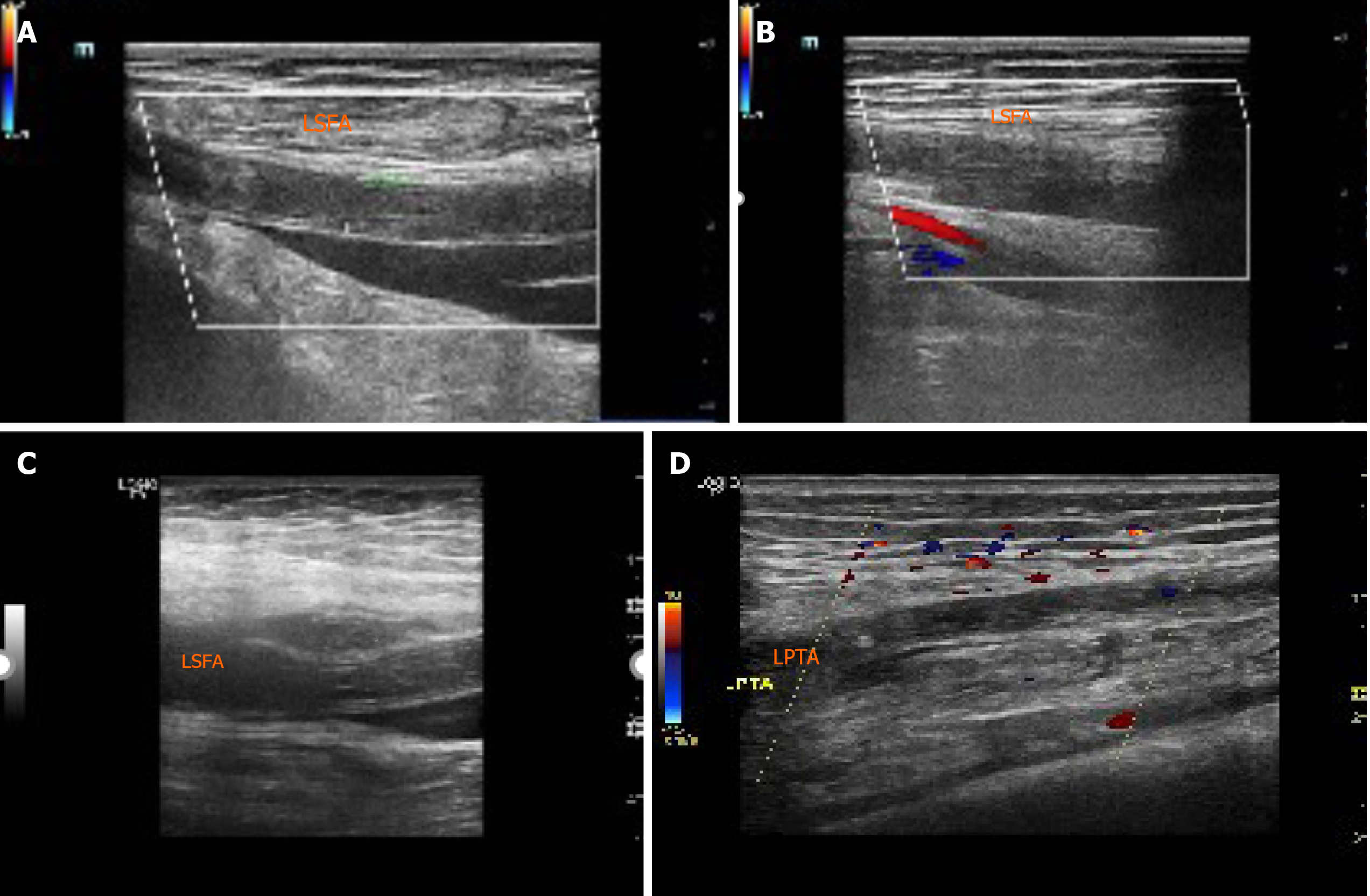
Figure 1 Ultrasonographic findings.
A: Acute occlusion of the middle and upper segments of the left superficial femoral artery (LSFA); B: Formation of lateral branches in the lower segment of the LSFA; C: Second acute occlusion of the LSFA; D: Acute occlusion of the left posterior tibial artery. LPTA: Left posterior tibial artery; LSFA: Left superficial femoral artery.
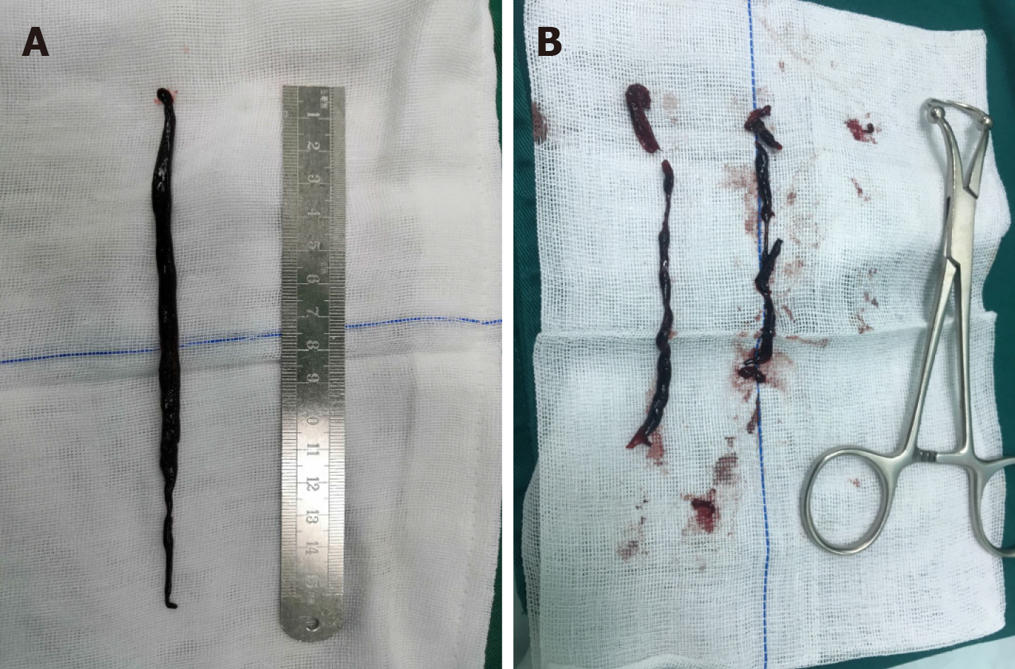
Figure 2 Thrombectomy photos.
A: A 15-cm long thrombus was removed during the first thrombectomy; B: A 10-cm long thrombus was removed during the second thrombectomy.
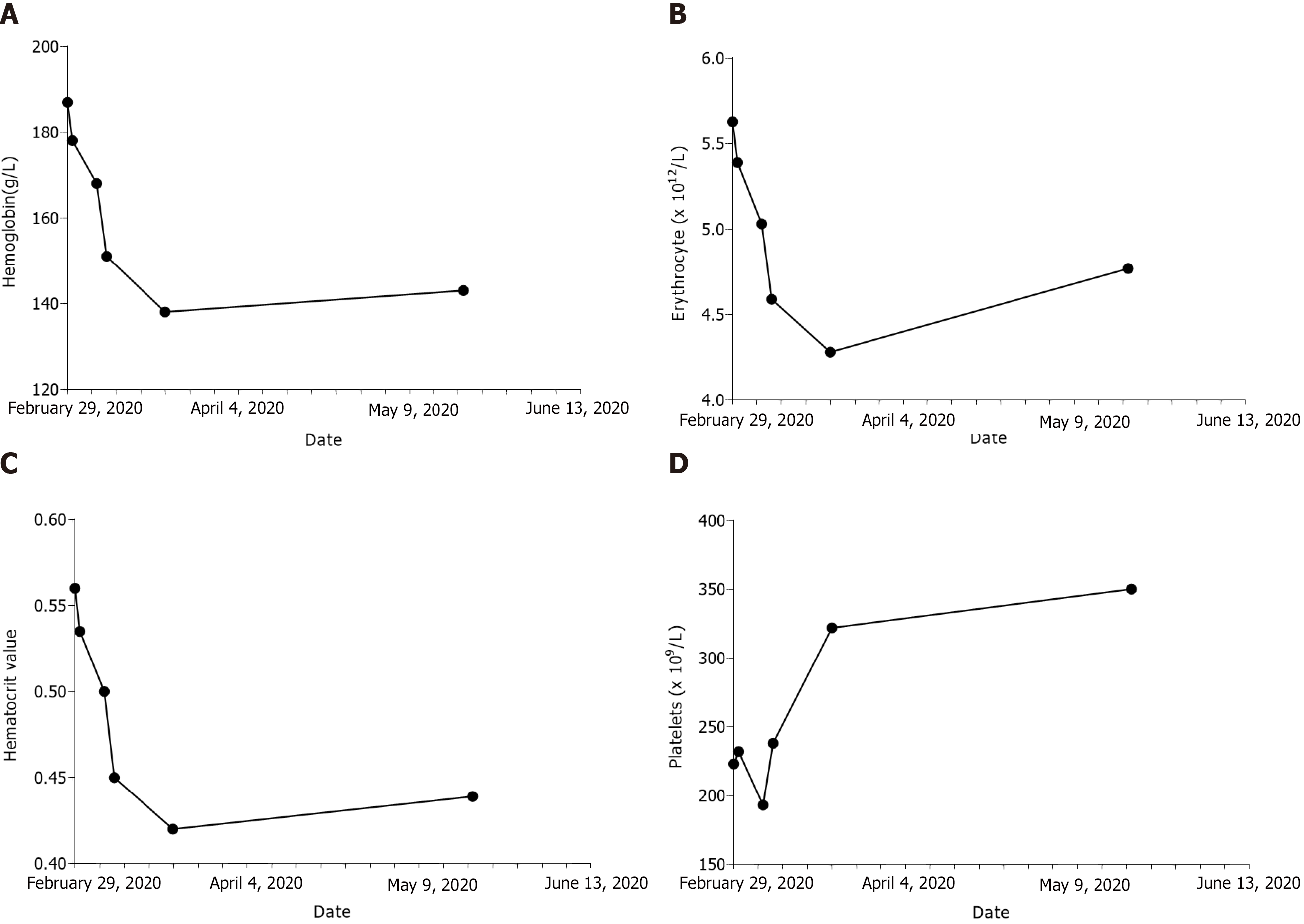
Figure 3 Change trends of relevant hematological indices during the perioperative period.
A: Hemoglobin; B: Erythrocyte; C: Hematocrit value; D: Platelets.
- Citation: Jiang BP, Cheng GB, Hu Q, Wu JW, Li XY, Liao S, Wu SY, Lu W. Recurrent thrombosis in the lower extremities after thrombectomy in a patient with polycythemia vera: A case report. World J Clin Cases 2020; 8(24): 6473-6479
- URL: https://www.wjgnet.com/2307-8960/full/v8/i24/6473.htm
- DOI: https://dx.doi.org/10.12998/wjcc.v8.i24.6473


