Copyright
©The Author(s) 2020.
World J Clin Cases. Dec 26, 2020; 8(24): 6432-6436
Published online Dec 26, 2020. doi: 10.12998/wjcc.v8.i24.6432
Published online Dec 26, 2020. doi: 10.12998/wjcc.v8.i24.6432
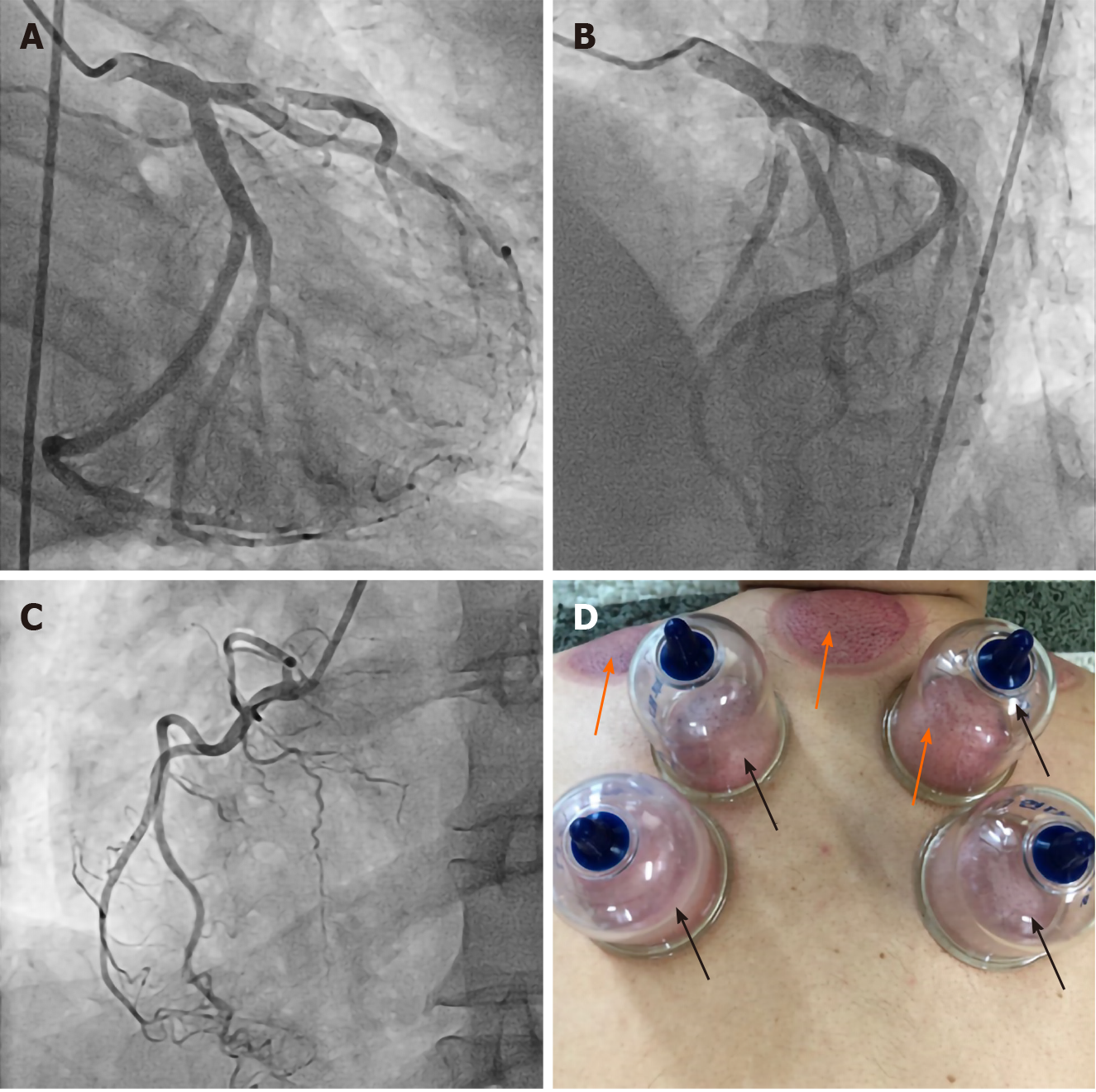
Figure 1 Images of coronary angiography and an illustration of wet cupping.
A-C: Coronary angiographic images of the left and right coronary arteries; A and B: A ruptured plaque is observed in the proximal left anterior descending artery; D: A picture of cupping; plastic cups (black arrows) are attached to skin using air suction to create subatmospheric pressure. Skin pricking precedes this process in wet cupping (WC), which facilitates bloodletting. Bruises (orange arrows) develop after WC.
- Citation: Jang AY, Suh SY. Extreme venous letting and cupping resulting in life-threatening anemia and acute myocardial infarction: A case report. World J Clin Cases 2020; 8(24): 6432-6436
- URL: https://www.wjgnet.com/2307-8960/full/v8/i24/6432.htm
- DOI: https://dx.doi.org/10.12998/wjcc.v8.i24.6432


