Copyright
©The Author(s) 2020.
World J Clin Cases. Dec 26, 2020; 8(24): 6346-6352
Published online Dec 26, 2020. doi: 10.12998/wjcc.v8.i24.6346
Published online Dec 26, 2020. doi: 10.12998/wjcc.v8.i24.6346
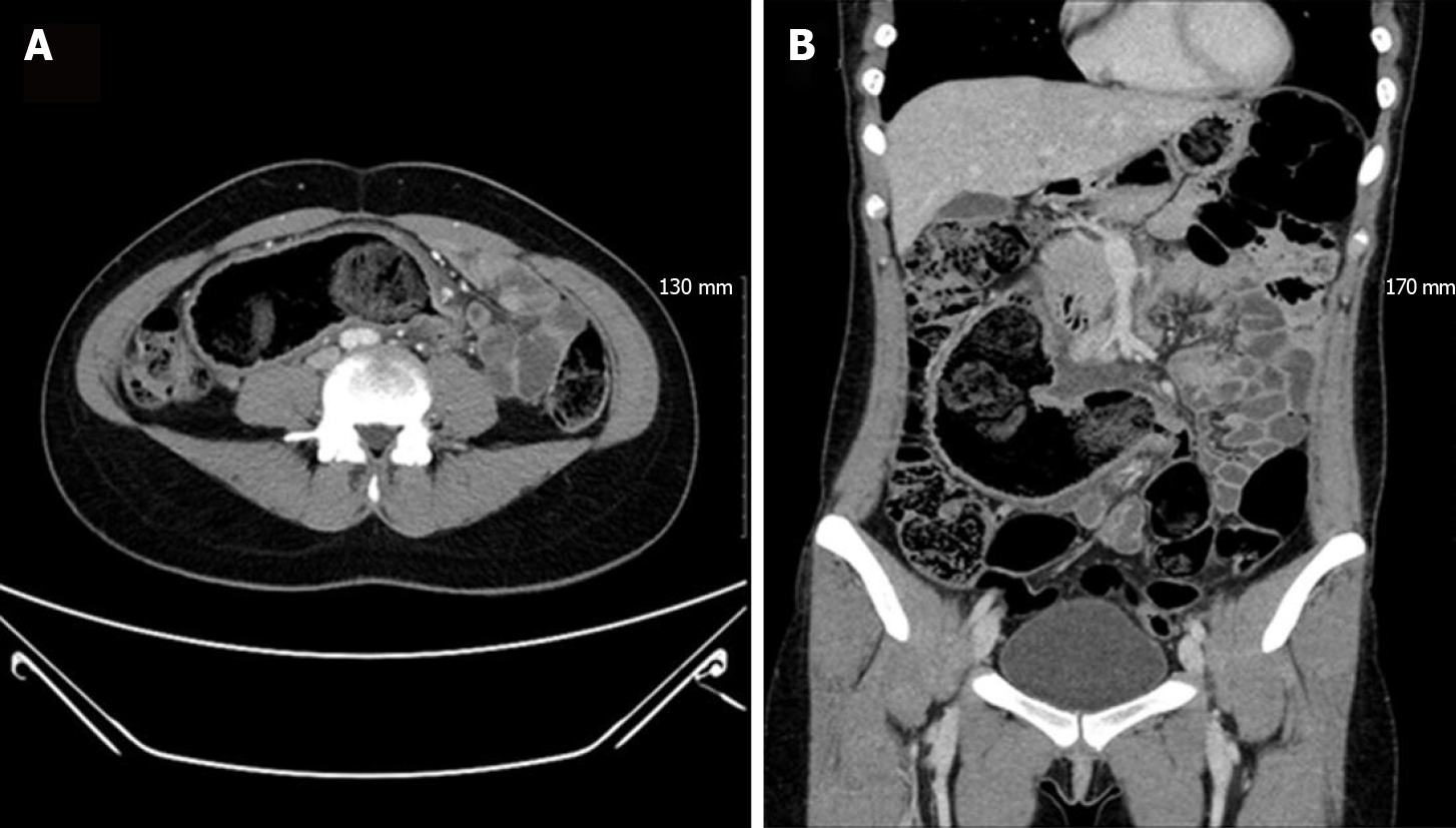
Figure 1 Abdomino-pelvic computed tomography showed a blind, dilated bowel loop filled with fecal material, directed to right upper quadrant of abdomen.
A: Axial view; B: Coronal view.
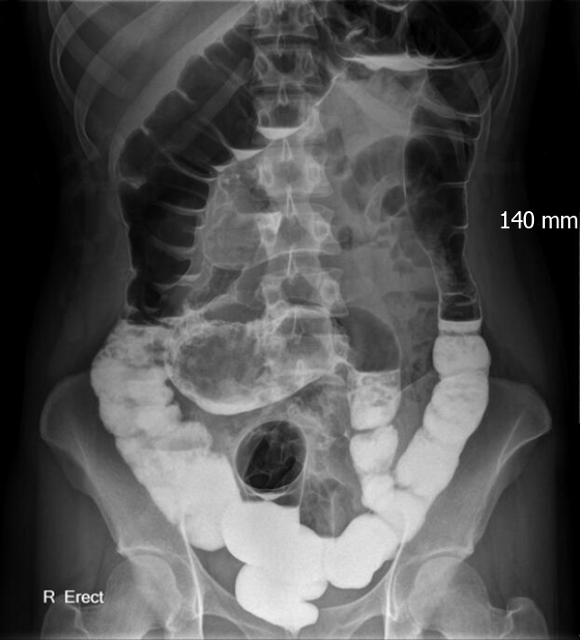
Figure 2 Gastrografin colon study revealed a Y-shaped structure formed by the sigmoid colon and duplicated colonic segment.
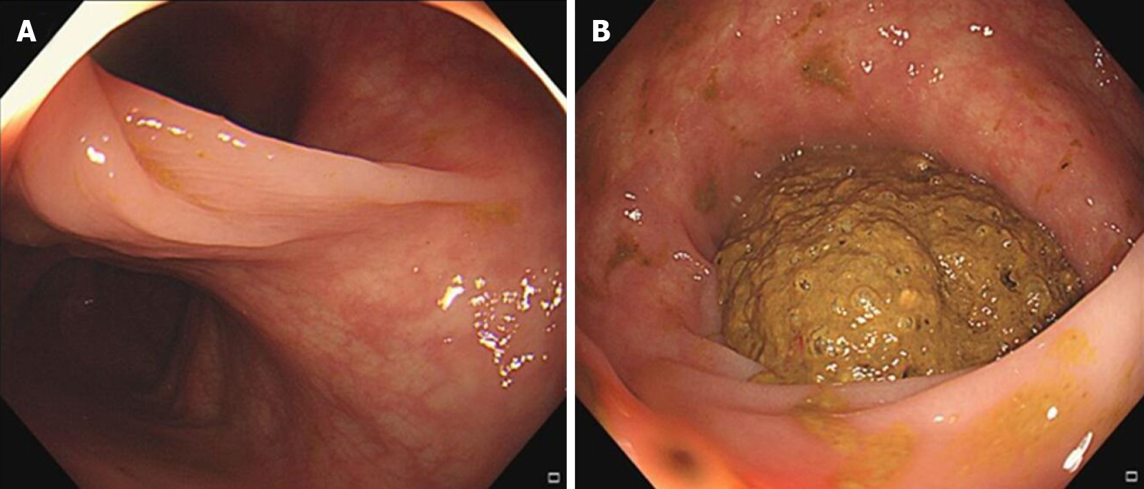
Figure 3 Colonoscopy images.
A: Bifurcation of the colonic lumen; B: The duplicated segment was filled with huge fecalomas.
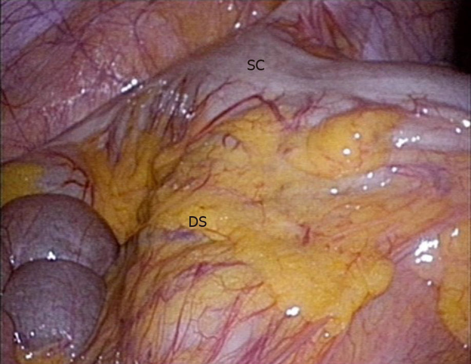
Figure 4 During the laparoscopic examinatin, an about 30 cm long, tubular bowel segment originating from the mesenteric side of the sigmoid colon was identified.
SC: Sigmoid colon; DS: Duplication segment.
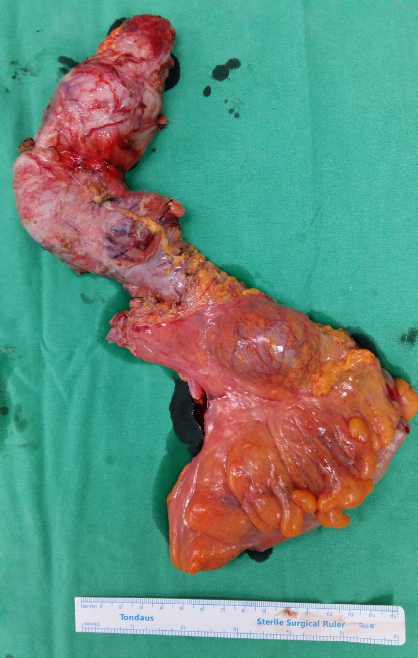
Figure 5 Grossly, the duplicated segment, measuring 34 cm in length, was connected to the native sigmoid colon at the mesenteric side.
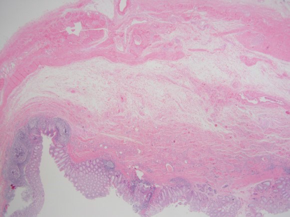
Figure 6 Microscopically, the duplication segment shows all 3 layers of bowel wall with scatted heterotopic tissue (Hematoxylin and eosin stain, ×12.
5).
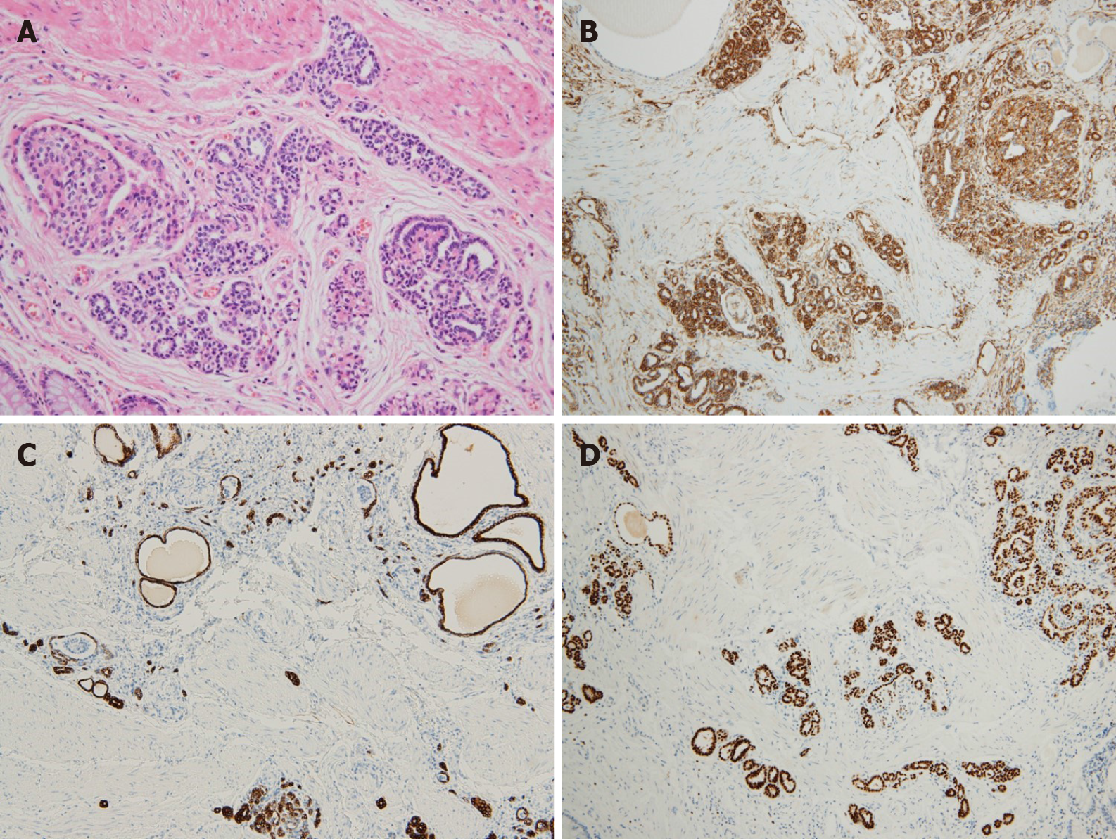
Figure 7 The heterotopic tissue is consistent with ectopic immature renal tissue.
A: Higher magnifications view (Hematoxylin and eosin stain, × 200); B: Immunohistochemical staining (× 200) for vimentin; C: Immunohistochemical staining (× 200) for CK7; D: Immunohistochemical staining (× 200) for PAX8.
- Citation: Namgung H. Sigmoid colon duplication with ectopic immature renal tissue in an adult: A case report. World J Clin Cases 2020; 8(24): 6346-6352
- URL: https://www.wjgnet.com/2307-8960/full/v8/i24/6346.htm
- DOI: https://dx.doi.org/10.12998/wjcc.v8.i24.6346


