Copyright
©The Author(s) 2020.
World J Clin Cases. Dec 26, 2020; 8(24): 6330-6336
Published online Dec 26, 2020. doi: 10.12998/wjcc.v8.i24.6330
Published online Dec 26, 2020. doi: 10.12998/wjcc.v8.i24.6330
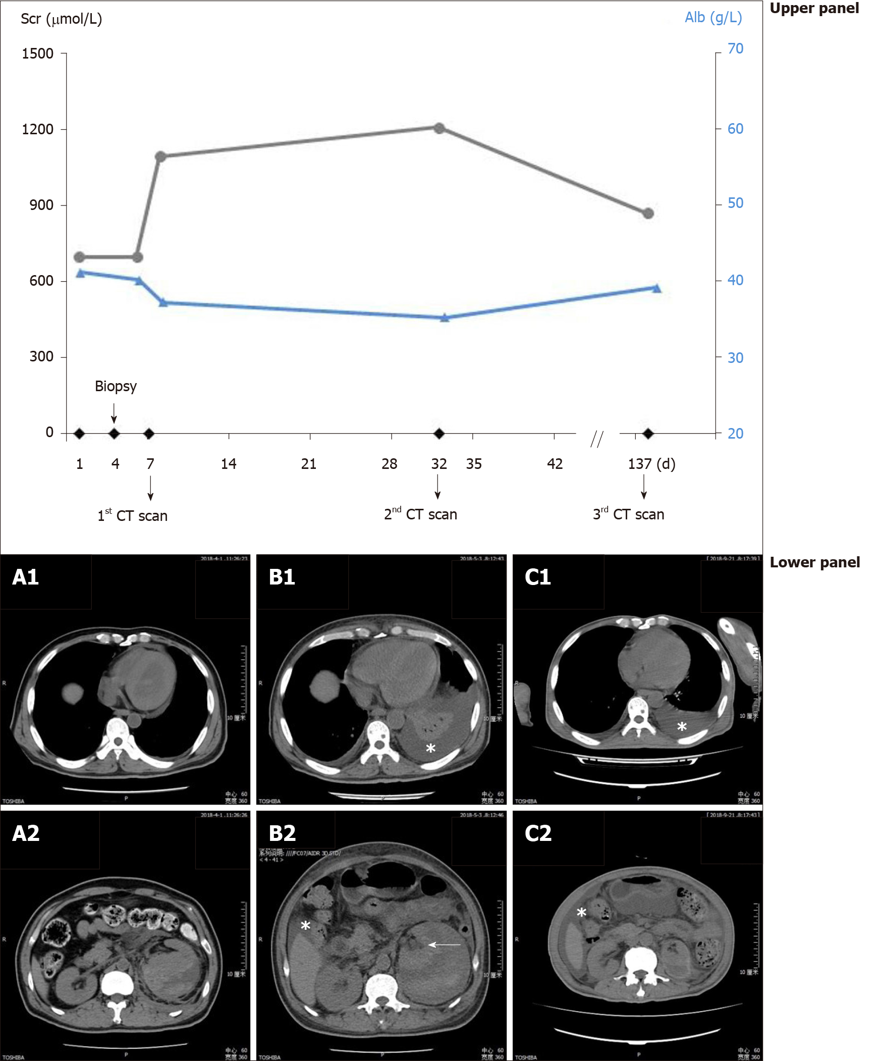
Figure 1 Evolution of the perirenal hematoma, pleural effusion and ascites in parallel with the corresponding serum creatinine and plasma albumin.
Upper panel: Values of serum creatinine (circles connected by black lines) and plasma albumin (triangles connected by blue lines) at different time points; Lower panel: A: Computed tomography scan shortly after the detection of hemorrhage showing perirenal hematoma (A2), without pleural effusion (A1); B: Perirenal hematoma and small amount of ascites (B2), with pleural effusion (B1, asterisk) 1 mo after the hemorrhage, the compressed kidney is also visible (B2, arrow); C: Perirenal fluid retention and ascites (C2), with pleural effusion (C1, asterisk) 5 mo after the hemorrhage. Alb: Albumin; CT: Computed tomography; Scr: Serum creatinine.
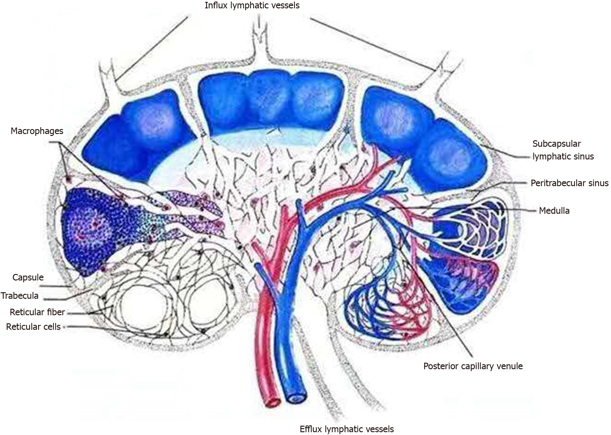
Figure 2 Schematic showing the flow of renal lymphatic fluid.
Inflow from the capsular lymph plexus went through the renal parenchyma via subcapsular and peritrabecular lymphatic sinuses and pericapillary space and eventually made confluence at the efflux lymphatic vessels for out-draining.
- Citation: Lin QZ, Wang HE, Wei D, Bao YF, Li H, Wang T. Pleural effusion and ascites in extrarenal lymphangiectasia caused by post-biopsy hematoma: A case report. World J Clin Cases 2020; 8(24): 6330-6336
- URL: https://www.wjgnet.com/2307-8960/full/v8/i24/6330.htm
- DOI: https://dx.doi.org/10.12998/wjcc.v8.i24.6330


