Copyright
©The Author(s) 2020.
World J Clin Cases. Dec 26, 2020; 8(24): 6322-6329
Published online Dec 26, 2020. doi: 10.12998/wjcc.v8.i24.6322
Published online Dec 26, 2020. doi: 10.12998/wjcc.v8.i24.6322
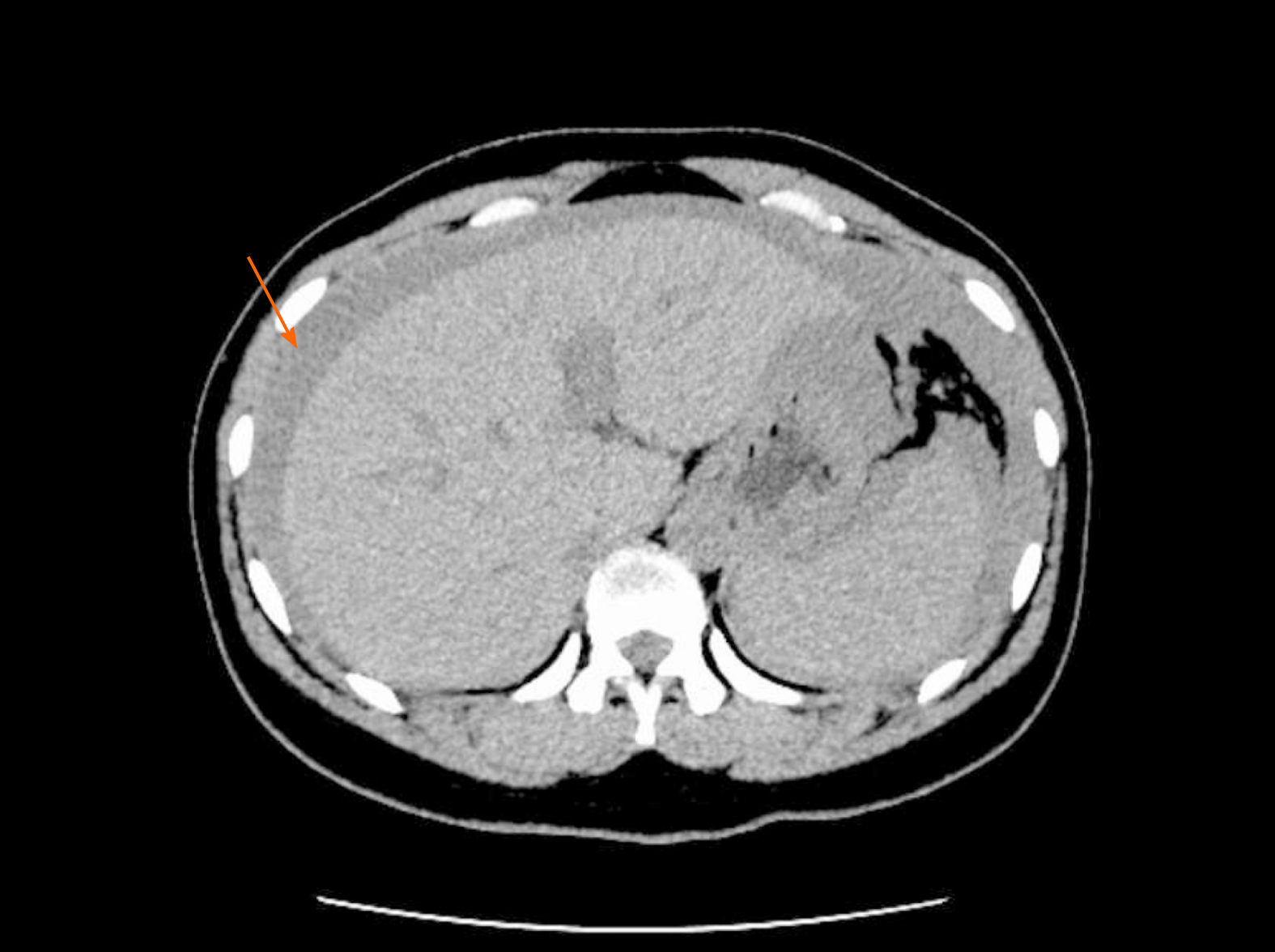
Figure 1 Computed tomography reveals pelvic and abdominal effusion.
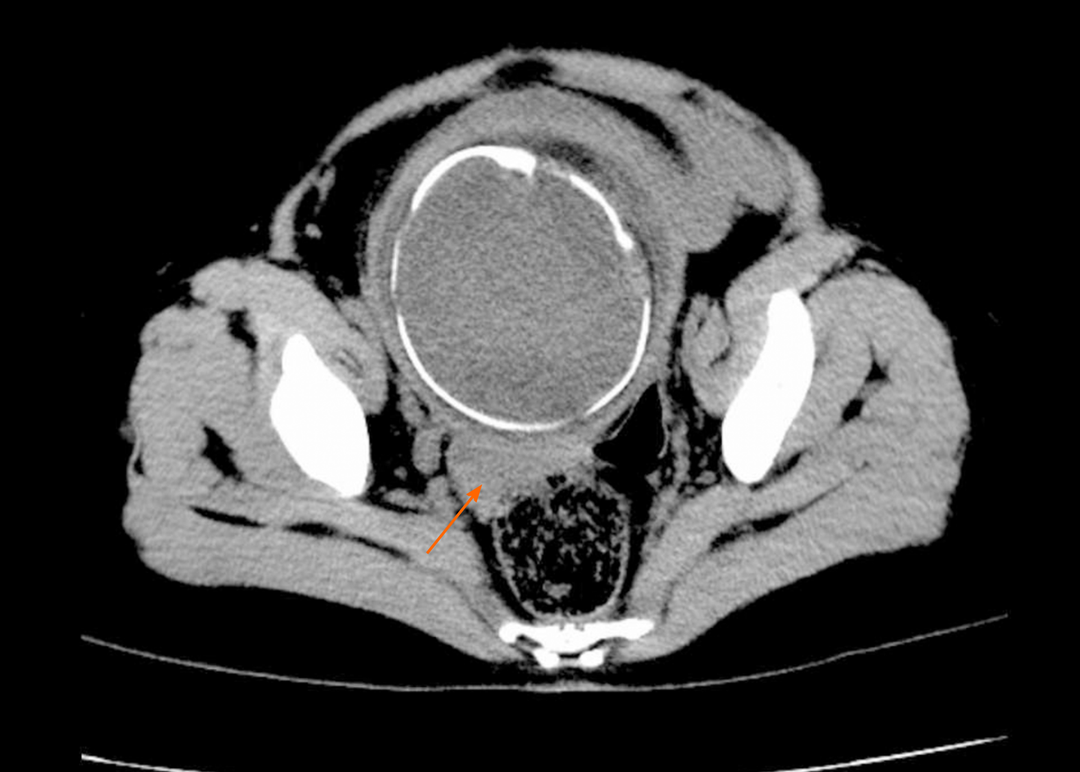
Figure 2 Computed tomography reveals a high probability of pelvic and abdominal hemoperitoneum.
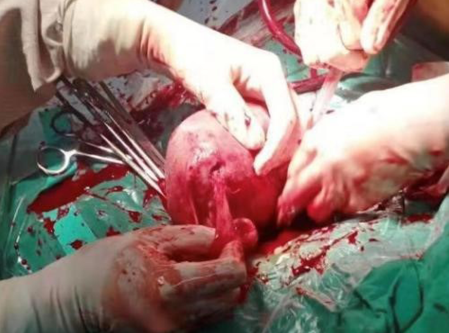
Figure 3 A rupture of 1.
5 cm is observed at the bottom of the left posterior wall of the uterus. The fimbrial portion of the left fallopian tube is completely adhered to the rupture.
Figure 4 A bulge (4 cm × 4 cm) is observed in the anterior wall of the right corner of the uterus, which is only composed of the serous layer and some weak muscle layers of the uterus.
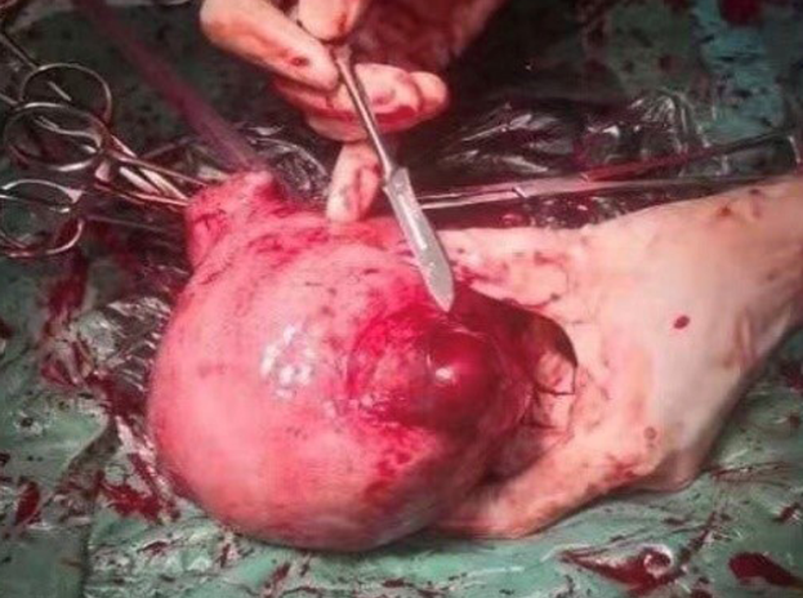
Figure 5 A 0.
5-cm rupture can be seen on the surface with active bleeding.
- Citation: Deng MF, Zhang XD, Zhang QF, Liu J. Uterine rupture in patients with a history of multiple curettages: Two case reports. World J Clin Cases 2020; 8(24): 6322-6329
- URL: https://www.wjgnet.com/2307-8960/full/v8/i24/6322.htm
- DOI: https://dx.doi.org/10.12998/wjcc.v8.i24.6322


