Copyright
©The Author(s) 2020.
World J Clin Cases. Dec 6, 2020; 8(23): 6206-6212
Published online Dec 6, 2020. doi: 10.12998/wjcc.v8.i23.6206
Published online Dec 6, 2020. doi: 10.12998/wjcc.v8.i23.6206
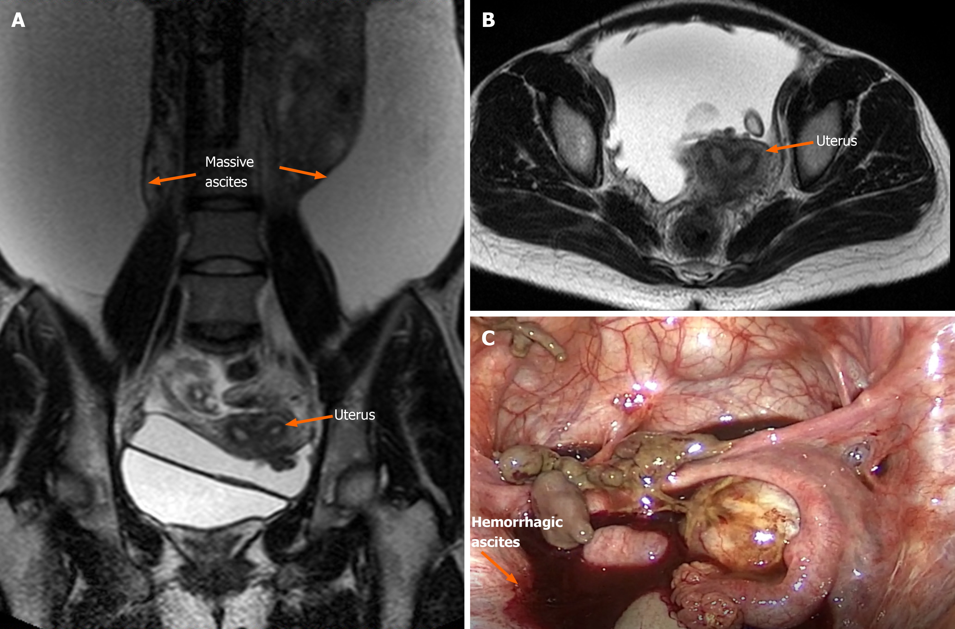
Figure 1 Bilateral ovaries were adhered to the posterior of the uterus, and endometriotic cysts were not observed in either ovary.
A and B: Magnetic resonance imaging revealed a large amount of ascites, and slight invagination of the fundus uteri, which proceeded to combine with bicornuate uterus; C: Large amount of brown ascites and coagulated blood were observed in the pelvic cavity.
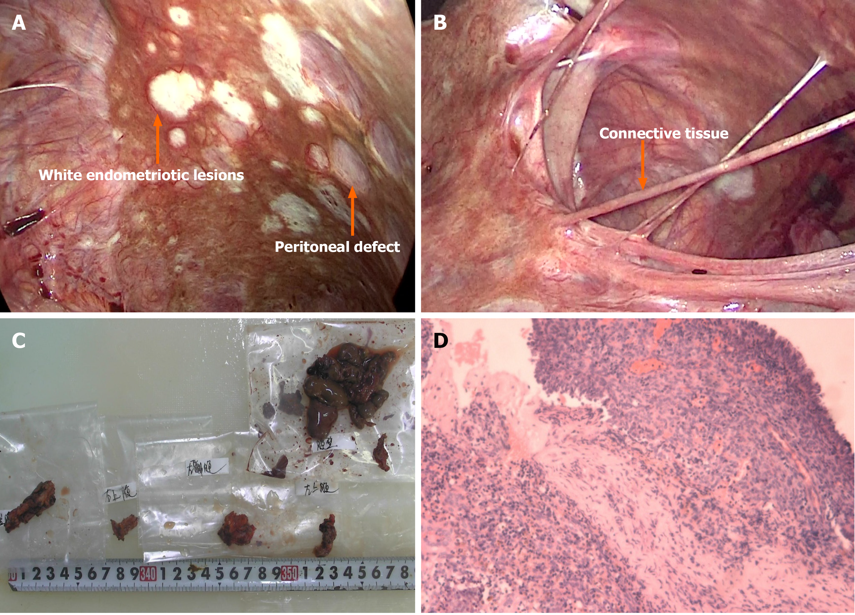
Figure 2 The final diagnosis.
A: The pelvic and abdominal peritonea were covered with patchy red, white, and brown endometriotic lesions and defects; B: All surfaces of abdominal organs were covered with tough connective tissue; C: Gross pathology of resected tissues; D: The final pathological diagnosis was endometriosis coupled with hemorrhagic necrotic tissue.
- Citation: Han X, Zhang ST. Novel triple therapy for hemorrhagic ascites caused by endometriosis: A case report. World J Clin Cases 2020; 8(23): 6206-6212
- URL: https://www.wjgnet.com/2307-8960/full/v8/i23/6206.htm
- DOI: https://dx.doi.org/10.12998/wjcc.v8.i23.6206


