Copyright
©The Author(s) 2020.
World J Clin Cases. Dec 6, 2020; 8(23): 6190-6196
Published online Dec 6, 2020. doi: 10.12998/wjcc.v8.i23.6190
Published online Dec 6, 2020. doi: 10.12998/wjcc.v8.i23.6190
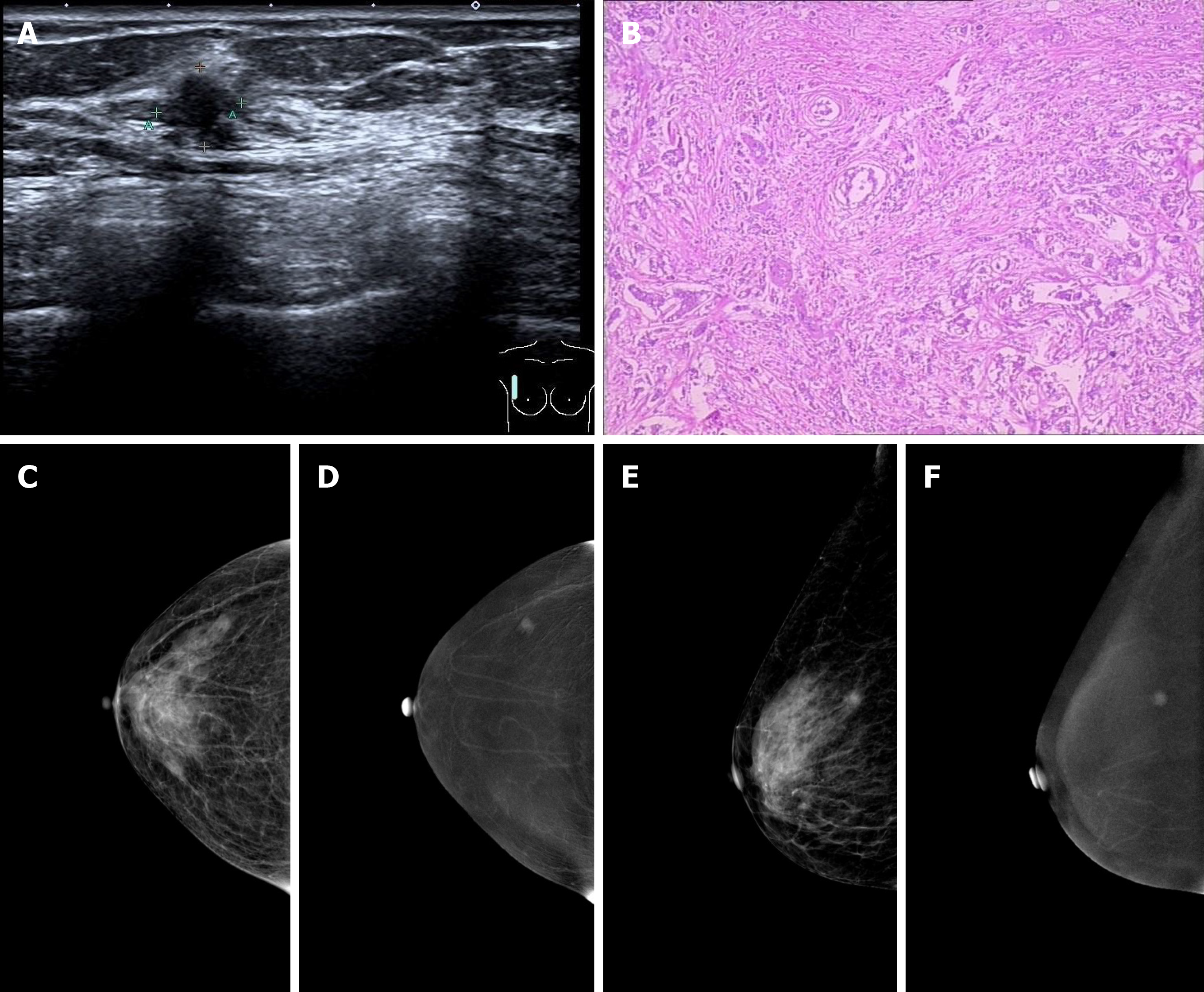
Figure 1 Breast imaging.
A: Ultrasound examination revealed an ill-defined hypoechoic mass in the right breast with an irregular shape; B: Microscopic examination revealed invasive ductal carcinoma cells invading the parenchyma in the right breast; C and E: On contrast enhanced spectral mammography (CESM) imaging, the low energy images showed the mass on the upper outer quadrant of the right breast with small spicules at the periphery; D and F: On CESM imaging, the subtraction images showed the significant enhancement of the lesions.
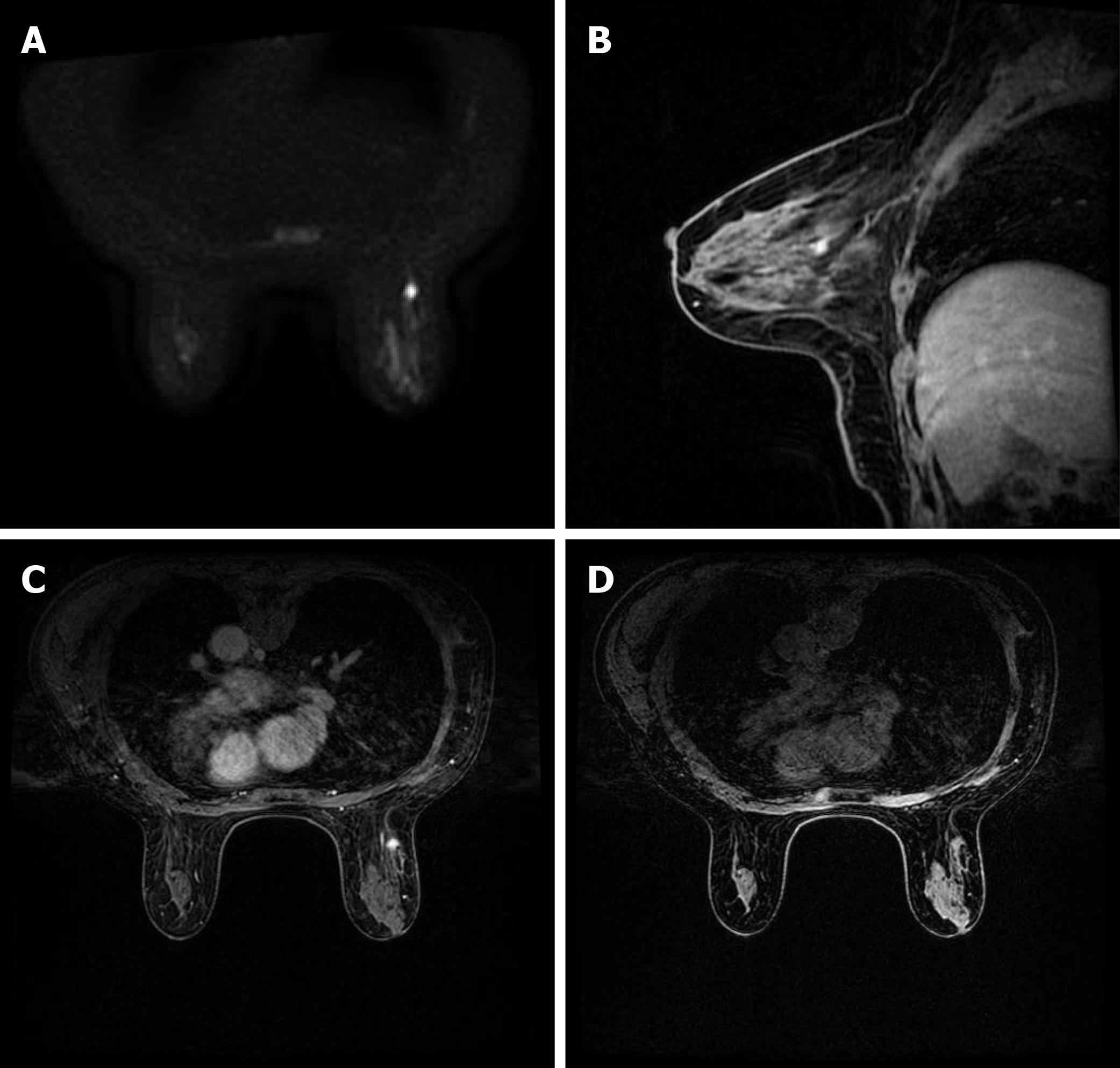
Figure 2 Brest imaging.
A: Axial diffusion weighted imaging (b = 800) showed the mass with high signal intensity; B and C: Sagittal and axial post-contrast fat-saturated T1-weighted magnetic resonance imaging identified a 0.8 × 0.9 cm enhancing irregular mass at the 9-10 o'clock position, approximately 6 cm from the nipple; D: Transverse non-enhanced T1 weighted imaging revealed an ill-defined mass with uniform equal signal intensity.
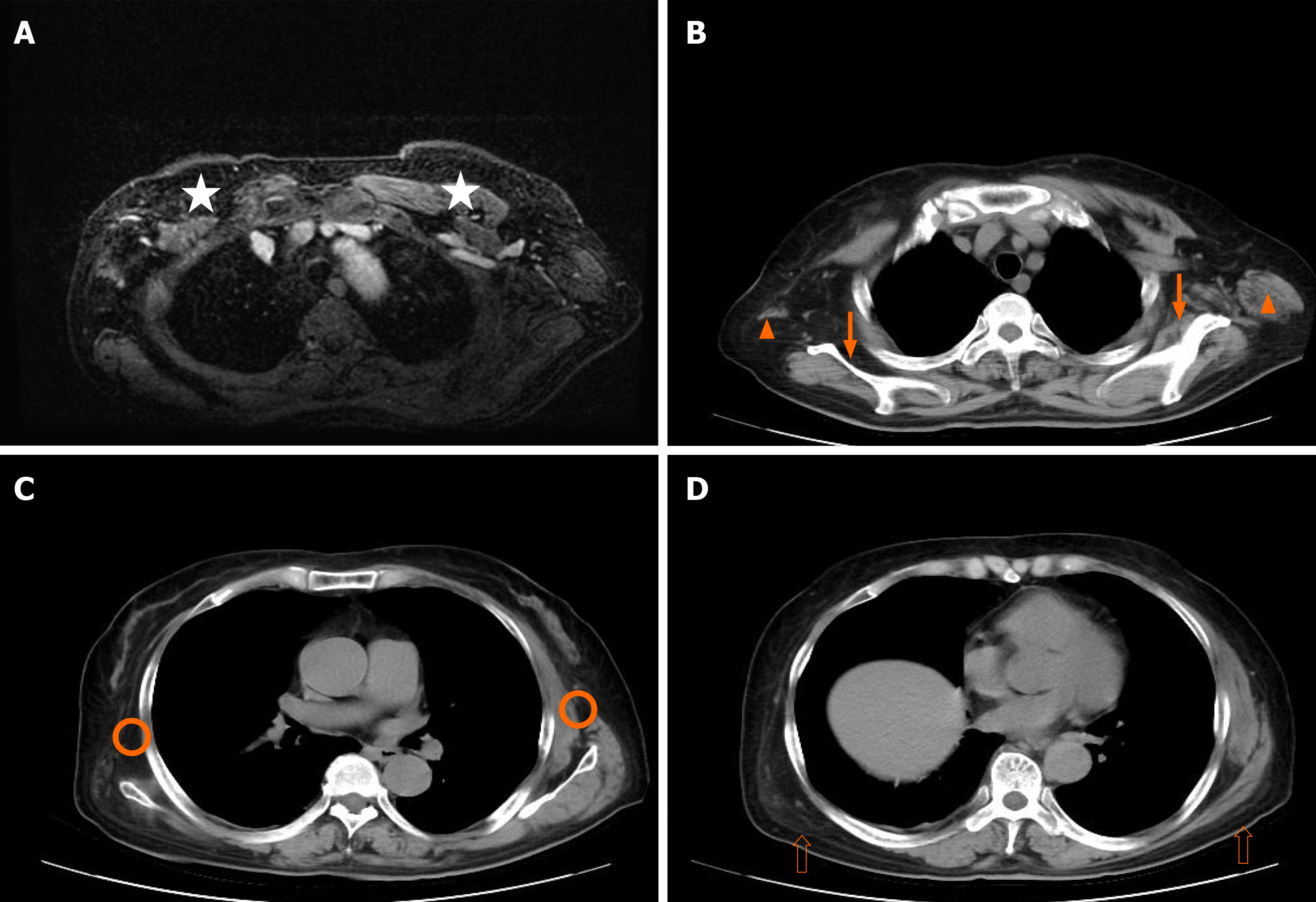
Figure 3 Chest imaging.
A: Axial magnetic resonance imaging; B, C, and D: CT scan of the chest revealed atrophy of the pectoralis major (star), teres major (orange triangle), serratus anterior (orange circle), subscapularis muscle (solid arrow), and latissimus dorsi muscle (hollow arrow).
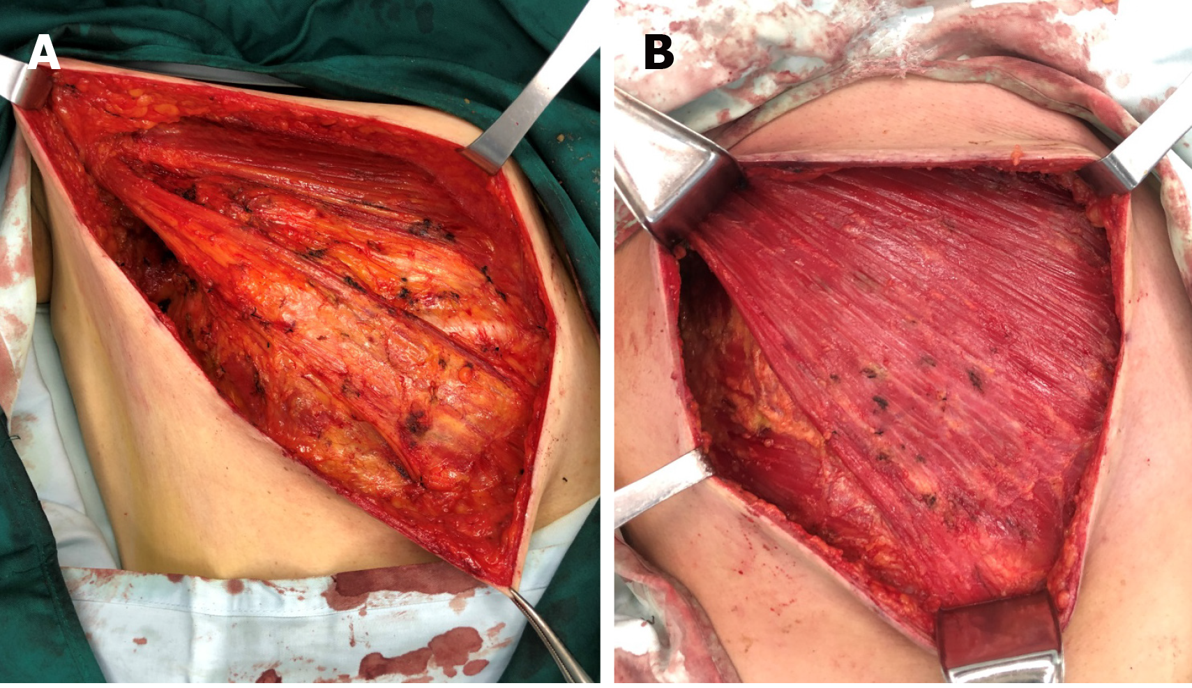
Figure 4 Breast surgery.
A: Pectoralis major, pectoralis minor, and serratus anterior muscles were atrophied, the muscle bundles were thin, and the boundary between the pectoralis major and fat and fascia of the posterior space of breast was ill-defined; B: Normal pectoralis major, pectoralis minor, and serratus anterior.
- Citation: Wang XM, Cong YZ, Qiao GD, Zhang S, Wang LJ. Primary breast cancer patient with poliomyelitis: A case report. World J Clin Cases 2020; 8(23): 6190-6196
- URL: https://www.wjgnet.com/2307-8960/full/v8/i23/6190.htm
- DOI: https://dx.doi.org/10.12998/wjcc.v8.i23.6190


