Copyright
©The Author(s) 2020.
World J Clin Cases. Dec 6, 2020; 8(23): 6150-6157
Published online Dec 6, 2020. doi: 10.12998/wjcc.v8.i23.6150
Published online Dec 6, 2020. doi: 10.12998/wjcc.v8.i23.6150
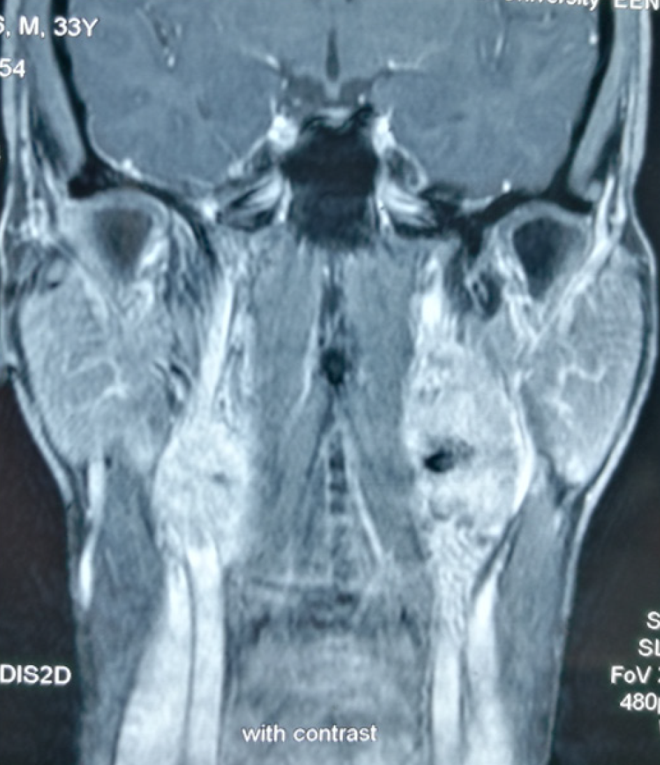
Figure 1 Magnetic resonance imaging showing bilateral carotid body tumors.
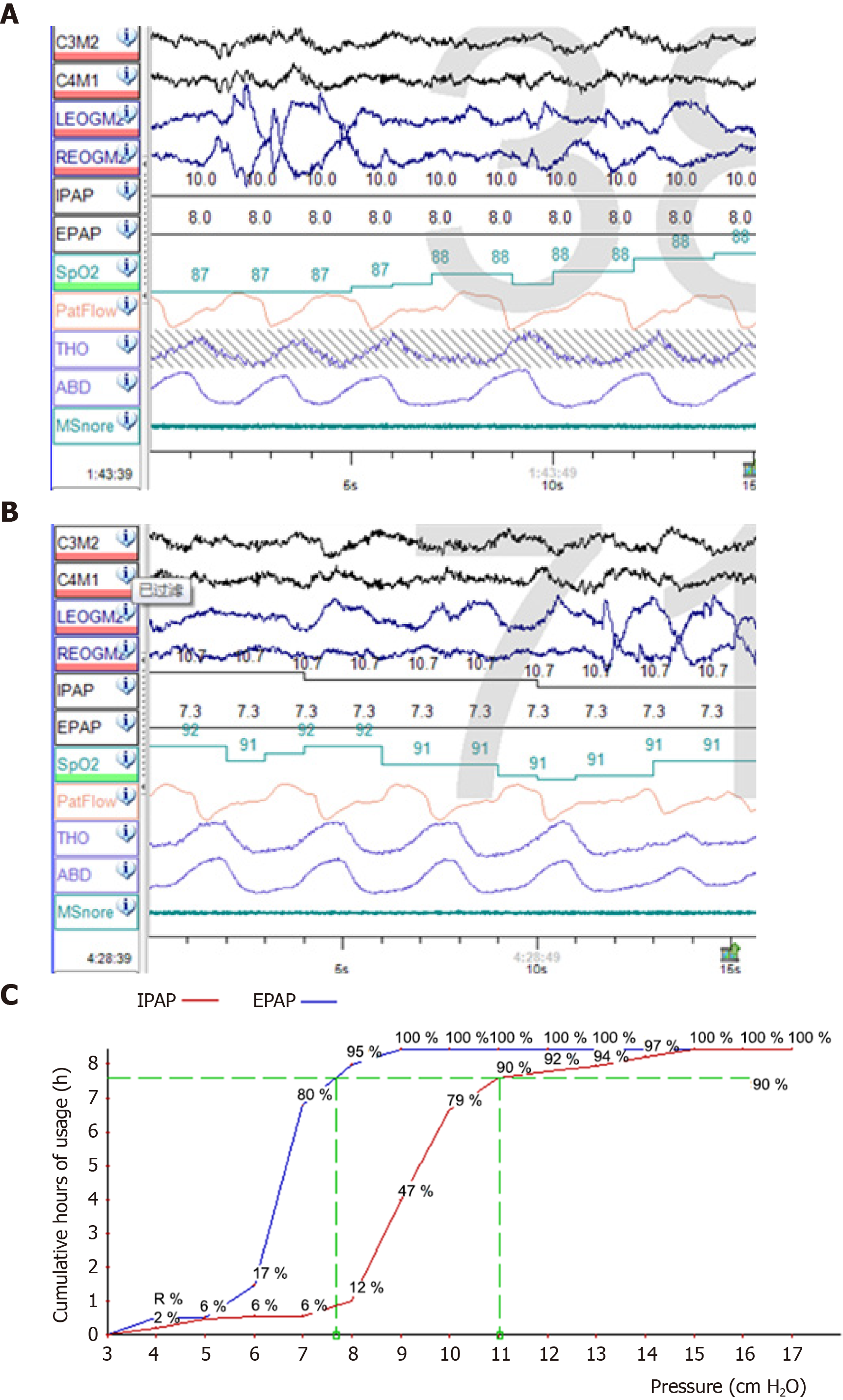
Figure 2 Manual pressure titration and pressure profile of the whole night manual pressure titration.
A: Manual pressure titration before oxygen therapy: During the whole-night pressure titration, at 1:43 am, the patient had no respiratory events or snoring, but saturation of pulse oximetry (SpO2) fluctuated between 85% and 88%; B: Manual pressure titration after oxygen therapy: Oxygen therapy at 1 L/min was given at 4:28 am. He reported no respiratory events or snoring during rapid eye movement sleep, and SpO2 fluctuated between 89% and 92%; C: Pressure profile of the whole night manual pressure titration: The whole-night pressure titration was 90% expiratory positive airway pressure 7.5 cmH2O and 90% inspiratory positive airway pressure 11 cmH2O. Pressure titration achieved good effects. IPAP: Inspiratory positive airway pressure; EPAP: expiratory positive airway pressure.
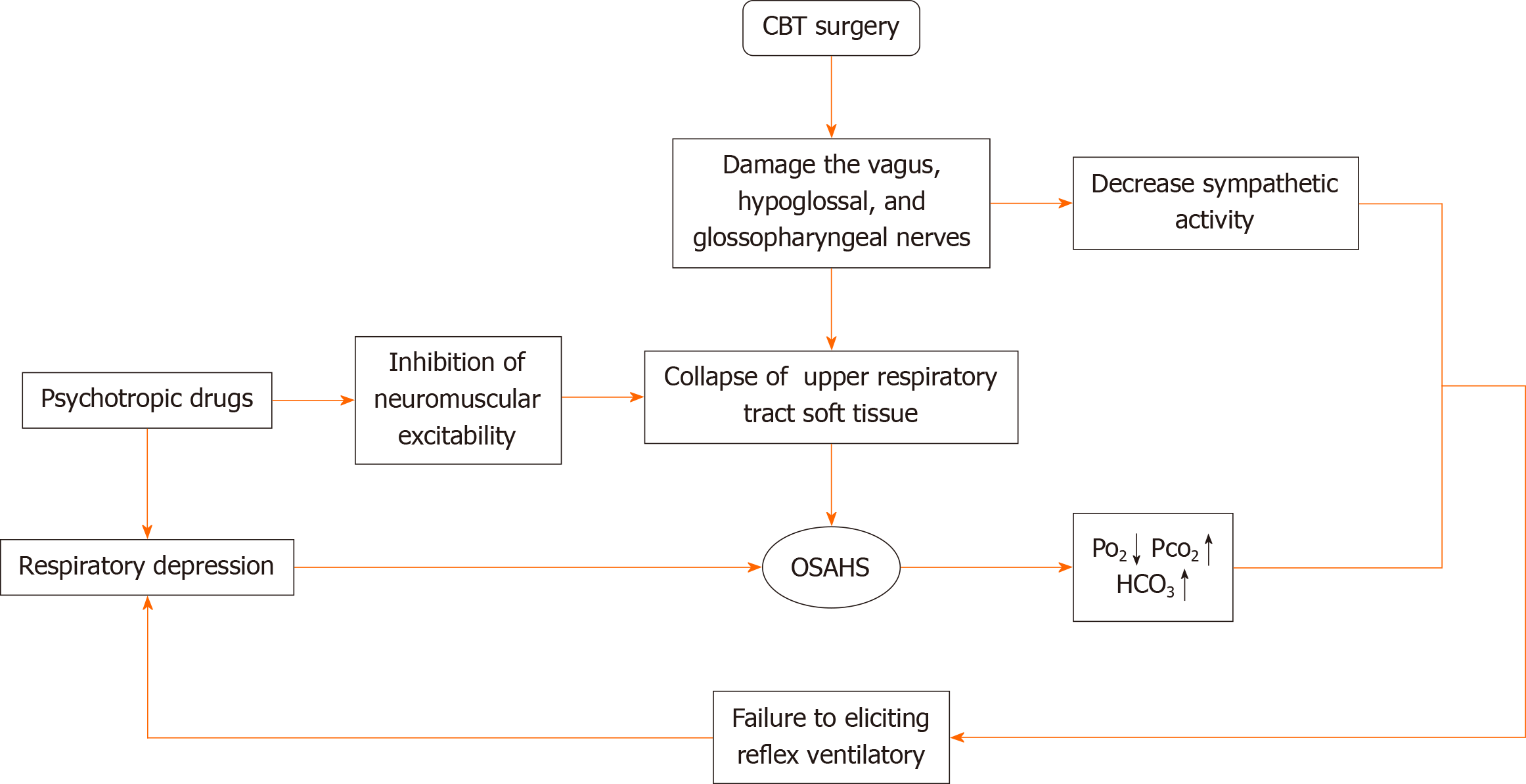
Figure 3 Flow chart illustrating the physiopathologic mechanism of the secondary aggravation of obstructive sleep apnea–hypopnea syndrome and hypoxemia.
CBT: Carotid body tumor; OSAHS: Obstructive sleep apnea–hypopnea syndrome; Po2: Oxygen pressure; Pco2: Carbon dioxide pressure; HCO3: Bicarbonate.
- Citation: Yang X, He XG, Jiang DH, Feng C, Nie R. Postoperative secondary aggravation of obstructive sleep apnea-hypopnea syndrome and hypoxemia with bilateral carotid body tumor: A case report. World J Clin Cases 2020; 8(23): 6150-6157
- URL: https://www.wjgnet.com/2307-8960/full/v8/i23/6150.htm
- DOI: https://dx.doi.org/10.12998/wjcc.v8.i23.6150


