Copyright
©The Author(s) 2020.
World J Clin Cases. Dec 6, 2020; 8(23): 6110-6121
Published online Dec 6, 2020. doi: 10.12998/wjcc.v8.i23.6110
Published online Dec 6, 2020. doi: 10.12998/wjcc.v8.i23.6110
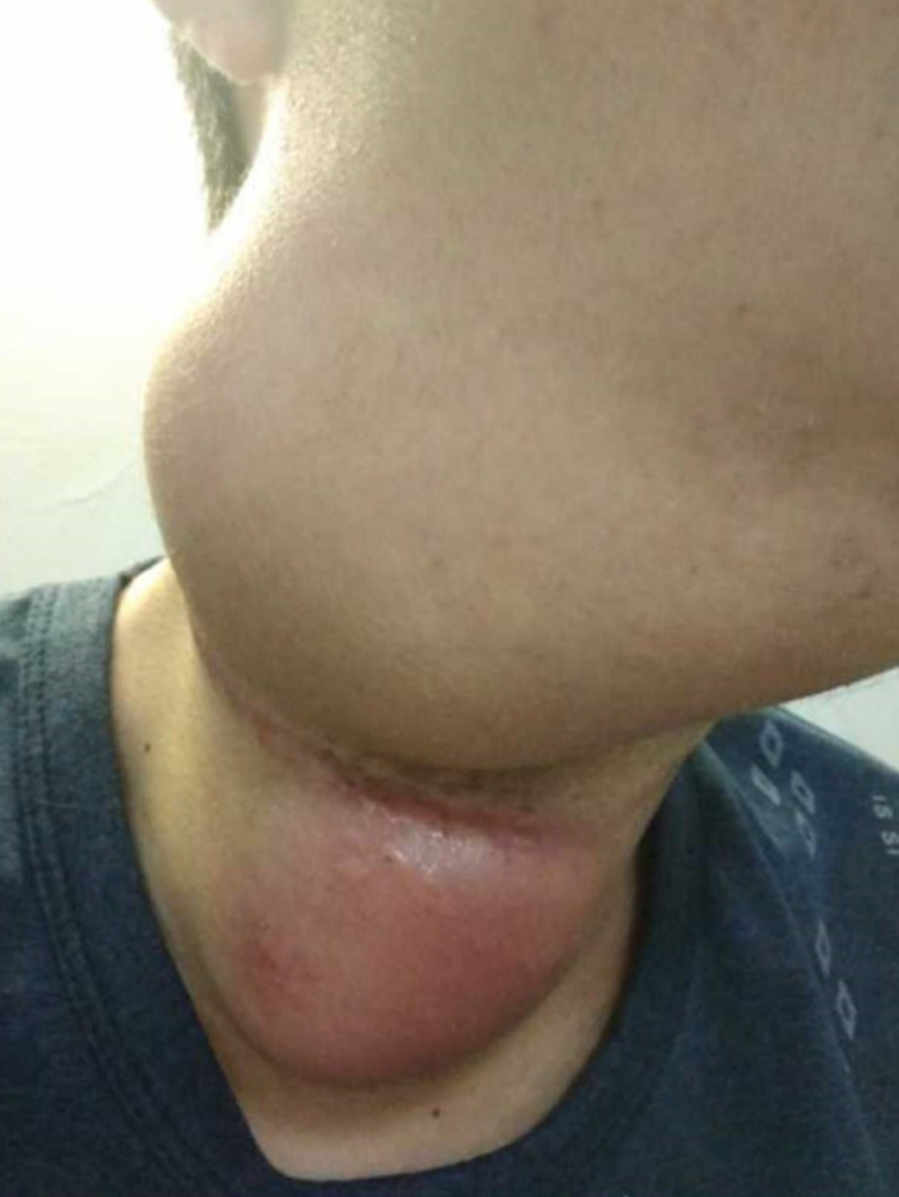
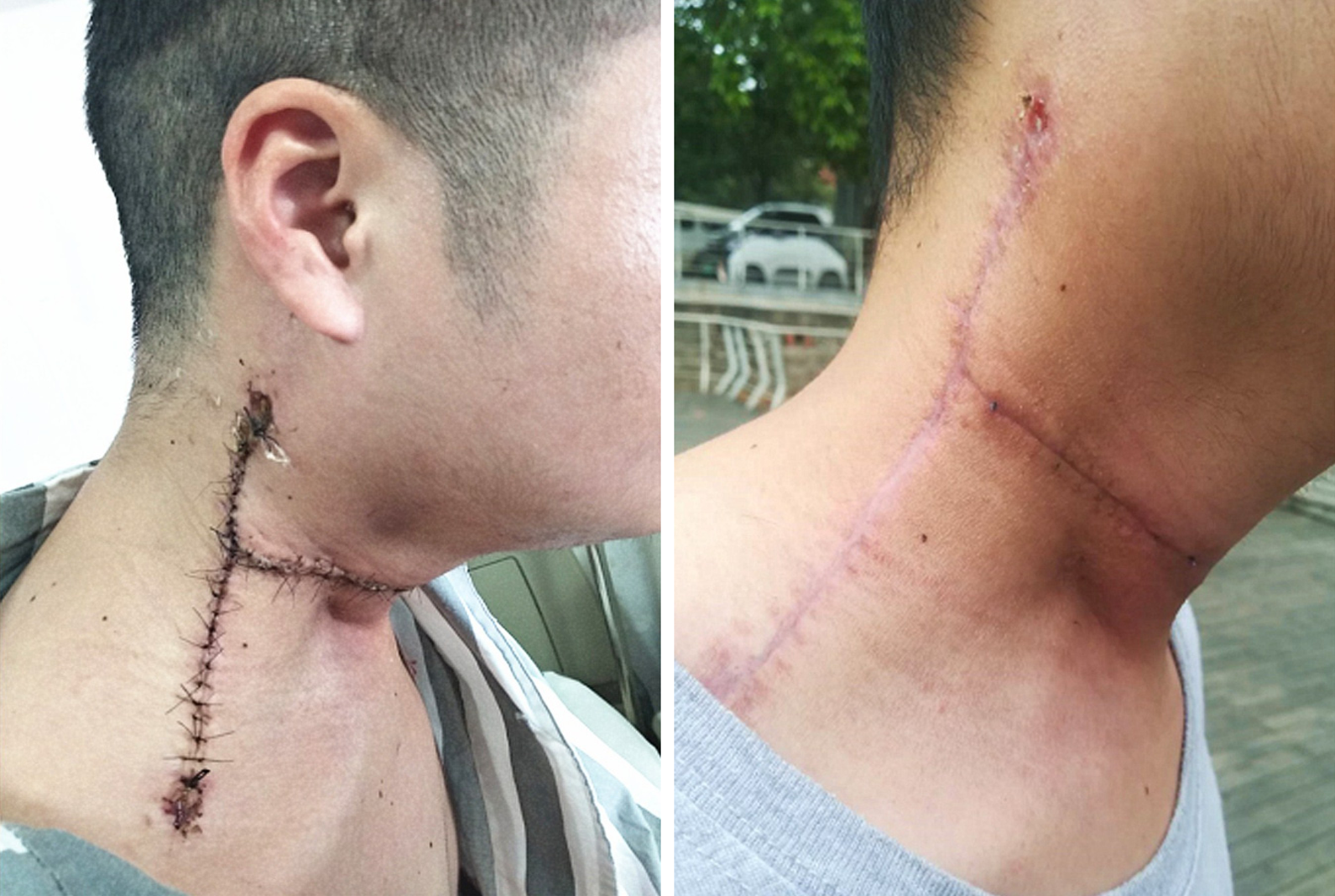
Figure 1 Appearance of the cervical masses.
Two masses in the upper and middle portions of the right neck, approximately 10 cm × 8 cm in the upper portion and 8 cm × 5 cm in the middle portion. An 18.0 cm lateral surgical scar was present between the two masses.
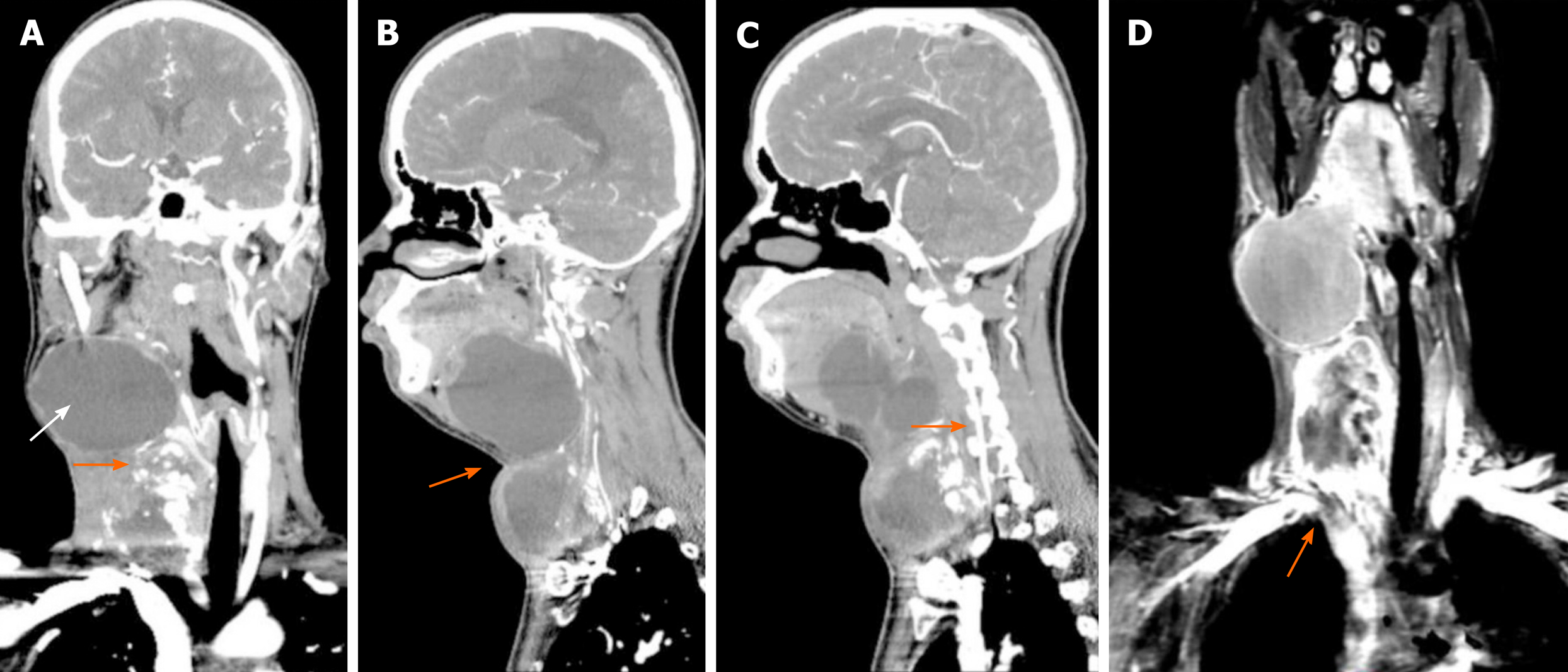
Figure 2 Computed tomography and magnetic resonance imaging.
A: Computed tomography imaging revealed a right side calcified mass and an unclear boundary at the root of the neck measuring 7.1 cm × 5.8 cm × 6.0 cm (orange arrow). Another large cystic mass was present in the right parapharyngeal space and measured 5.9 cm × 9.2 cm × 8.1 cm (white arrow). The two masses reached the right sublingual space and extended downward to the thorax. The adjacent blood vessels were displaced and the trachea, oropharynx, and hypopharynx were deviated to the left; B: The boundary between the upper and lower cervical masses is blurred (orange arrow); C: The vertebral artery is very close to the tumor (orange arrow). The vertebral artery runs inside the cervical spine. If the vertebral artery is injured during surgical resection of the tumor, it is likely to cause death due to the difficulty of hemostasis; D: Magnetic resonance imaging revealed a discontinuity of the angiography at the junction of the right subclavian artery (orange arrow) and the common carotid artery, indicating that the tumor has invaded the right subclavian artery.
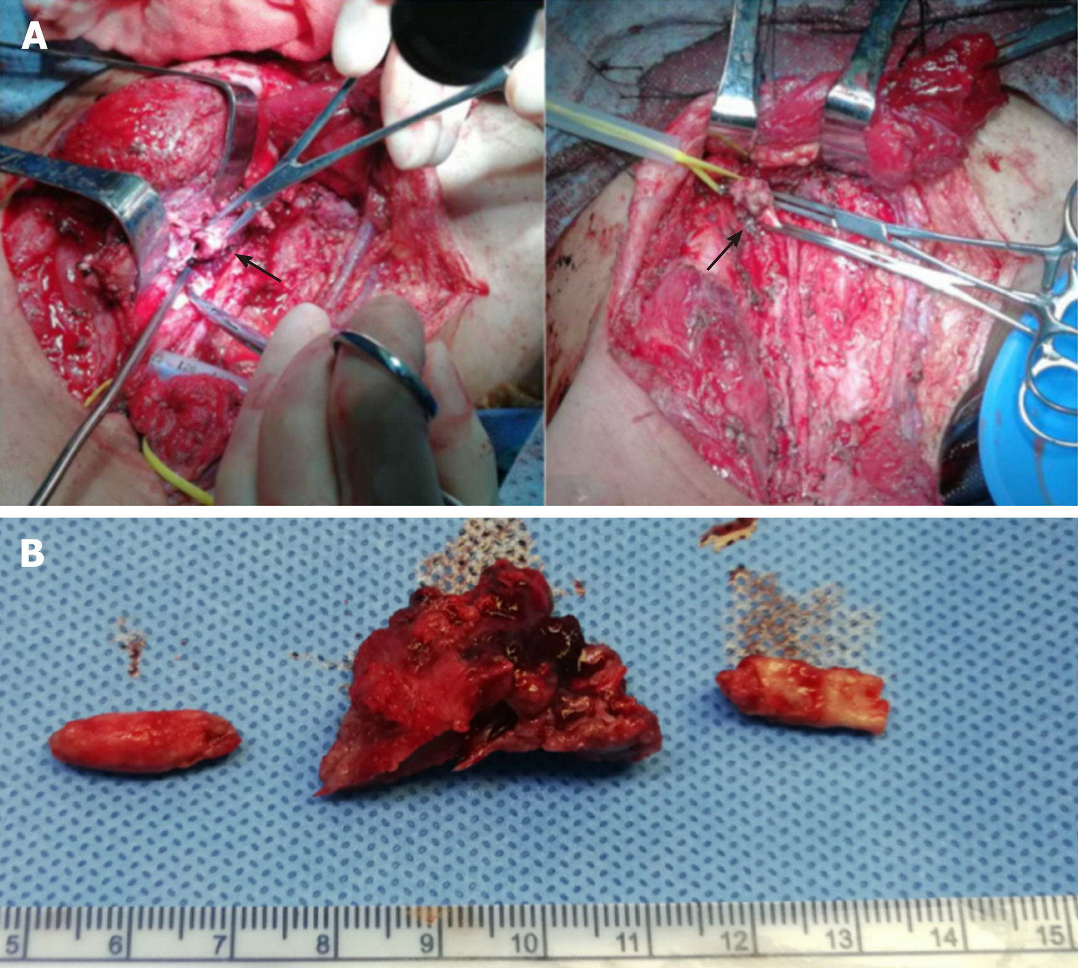
Figure 3 Surgical findings.
A: Shows a tumor thrombus in the right common carotid artery (left black arrow). Tumor invasion at the junction of the common carotid artery and the subclavian artery was also observed (right black arrow); B: Shows the removed tumor thrombus. The tumor thrombus in the artery is columnar in shape, the width of which is consistent with the inner diameter of the blood vessel, and there is a large amount of calcified tissue in the tumor.
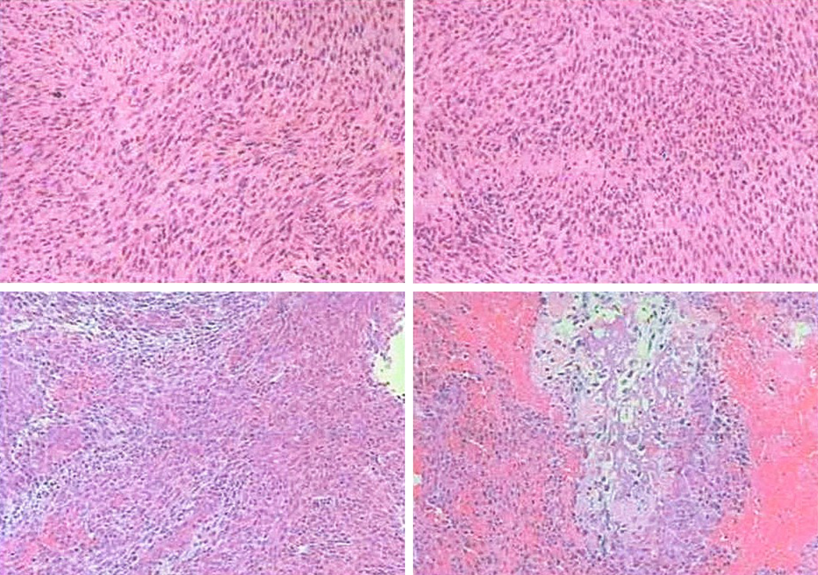
Figure 4 Microscopic pathology.
A spindle cell malignant tumor with local osteogenesis; and vimentin (+ ), CK (-), EMA (+), CK5 / 6 (-), P40 (-), CD34 (-), S-100 (-), SMA (+), TTF-1 (-), calponin [small focus] (+), desmin (-), PR (-), D2-40 (-), E-CAD (-), and Ki-67 [+ (50%)]. In situ hybridization showed EBERs (-). The following diagnoses were considered: (1) Osteosarcoma; (2) Malignant meningiomas with heterogeneous differentiation; (3) Tumor tissue noted in the common carotid artery; (4) Tumor tissue noted in the brachiocephalic trunk; and (5) One tracheoesophageal lymph node without metastasis.
Figure 5 Postoperative follow-up.
After surgery, the appearance of the neck had improved, and no signs of tumor were noted.
- Citation: Chen HY, Zhao F, Qin JY, Lin HM, Su JP. Malignant meningioma with jugular vein invasion and carotid artery extension: A case report and review of the literature. World J Clin Cases 2020; 8(23): 6110-6121
- URL: https://www.wjgnet.com/2307-8960/full/v8/i23/6110.htm
- DOI: https://dx.doi.org/10.12998/wjcc.v8.i23.6110


