Copyright
©The Author(s) 2020.
World J Clin Cases. Dec 6, 2020; 8(23): 6086-6094
Published online Dec 6, 2020. doi: 10.12998/wjcc.v8.i23.6086
Published online Dec 6, 2020. doi: 10.12998/wjcc.v8.i23.6086
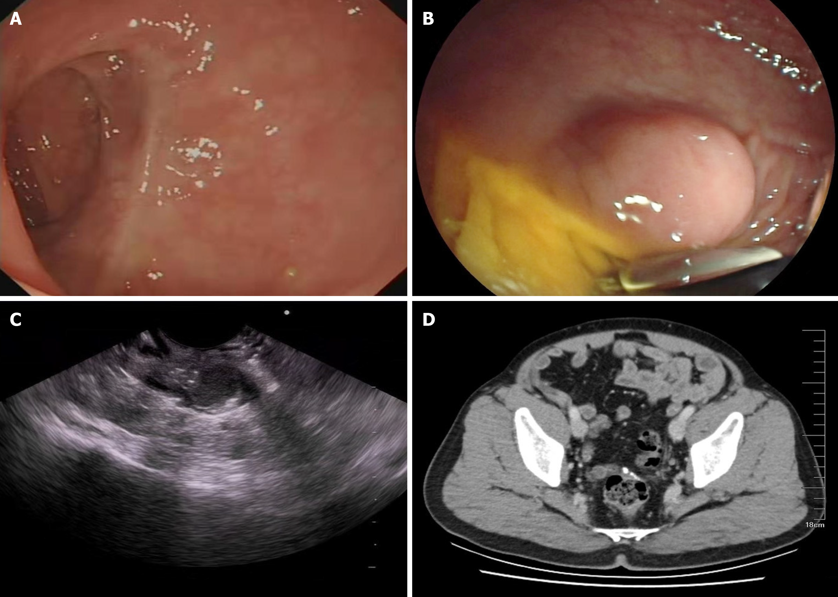
Figure 1 Anastomotic site findings.
A: Previous colonoscopy showing no abnormalities near the anastomotic site; B: Last colonoscopy performed at the local hospital showing a smooth protuberance near the anastomotic site; C: Endoscopic ultrasound image showing a hypoechoic structure in the rectum; D: No obvious thickening or strengthening of the anastomotic wall was visible in the contrast-enhanced computed tomography scan of the pelvic cavity.
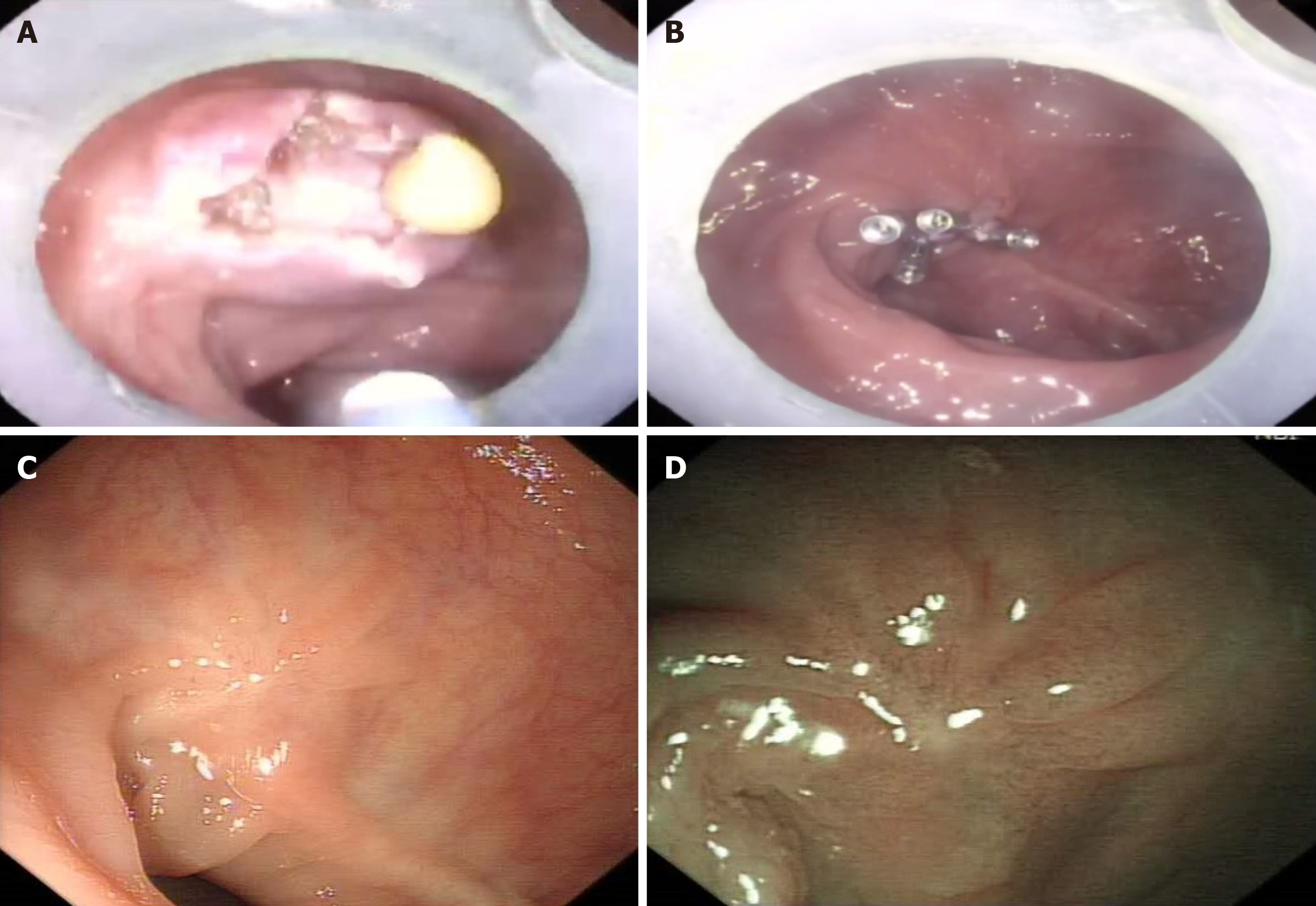
Figure 2 Endoscopic images.
A: After opening the sac wall, a yellow viscous liquid can be seen flowing out; B: Colonoscopy image showing the five metal clips clipping the wound after complete clearance of the abscess; C and D: There was no evidence of abscess recurrence in both white light endoscopy and narrow-band imaging after a follow-up period of 11 mo.
- Citation: Zhang BZ, Wang YD, Liao Y, Zhang JJ, Wu YF, Sun XL, Sun SY, Guo JT. Endoscopic fenestration in the diagnosis and treatment of delayed anastomotic submucosal abscess: A case report and review of literature. World J Clin Cases 2020; 8(23): 6086-6094
- URL: https://www.wjgnet.com/2307-8960/full/v8/i23/6086.htm
- DOI: https://dx.doi.org/10.12998/wjcc.v8.i23.6086


