Copyright
©The Author(s) 2020.
World J Clin Cases. Dec 6, 2020; 8(23): 6080-6085
Published online Dec 6, 2020. doi: 10.12998/wjcc.v8.i23.6080
Published online Dec 6, 2020. doi: 10.12998/wjcc.v8.i23.6080
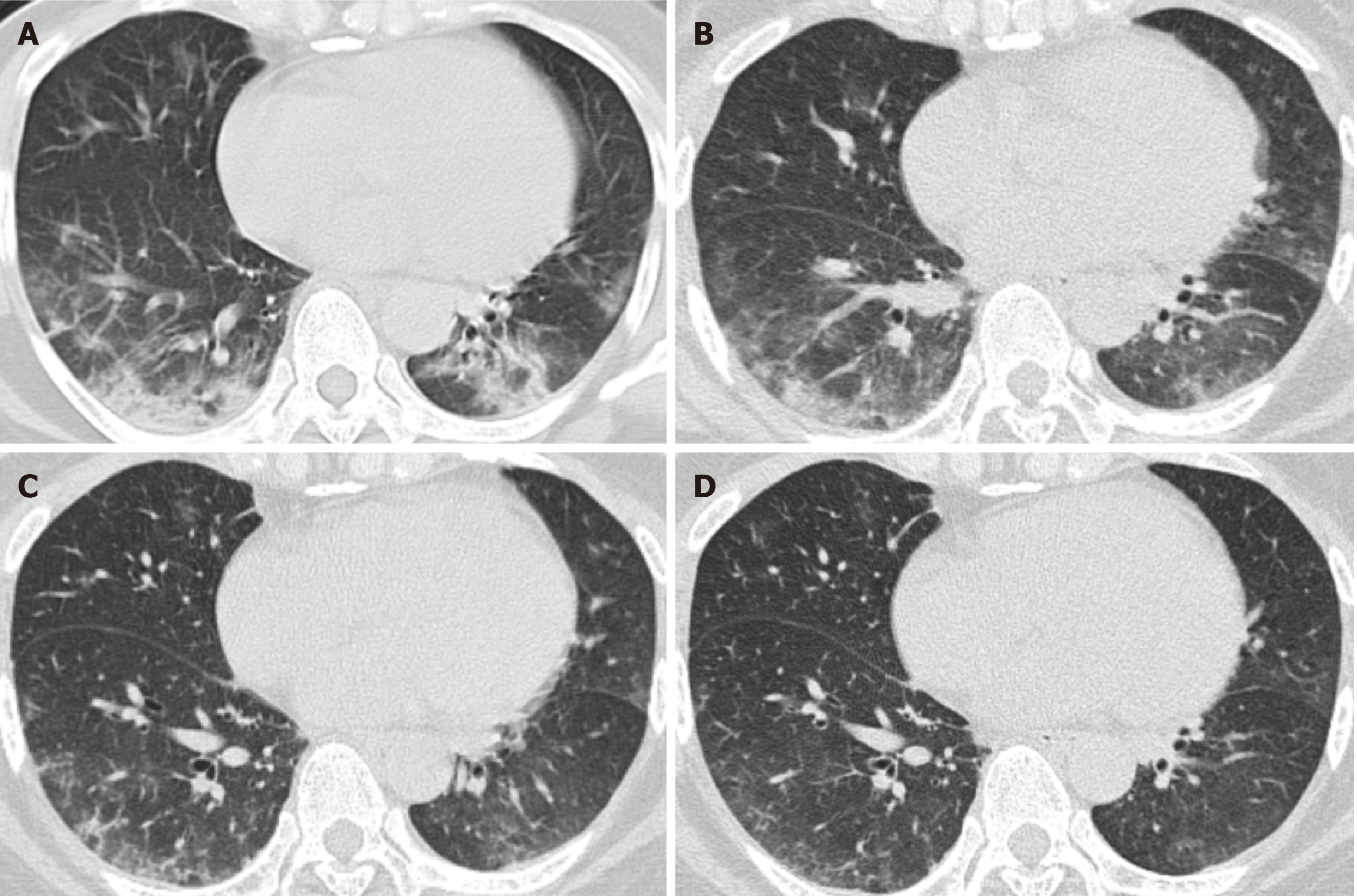
Figure 1 Unenhanced computed tomography images in a 49-year-old woman (axial imaging).
Computed tomography (CT) radiographs show multiple patchy ground-glass opacities and consolidated opacities in bilateral lower lobes, the middle lobe of the right lung and the tongue segment of the upper left lung, as treatment progressed, CT manifestations showed improvement. A: Day 7 (2020.1.26) after the onset of symptoms; B: Day 12 (2020.1.31) after the onset of symptoms; C: Day 16 (2020.2.4) after the onset of symptoms; D: Day 21 (2020.2.9) after the onset of symptoms.
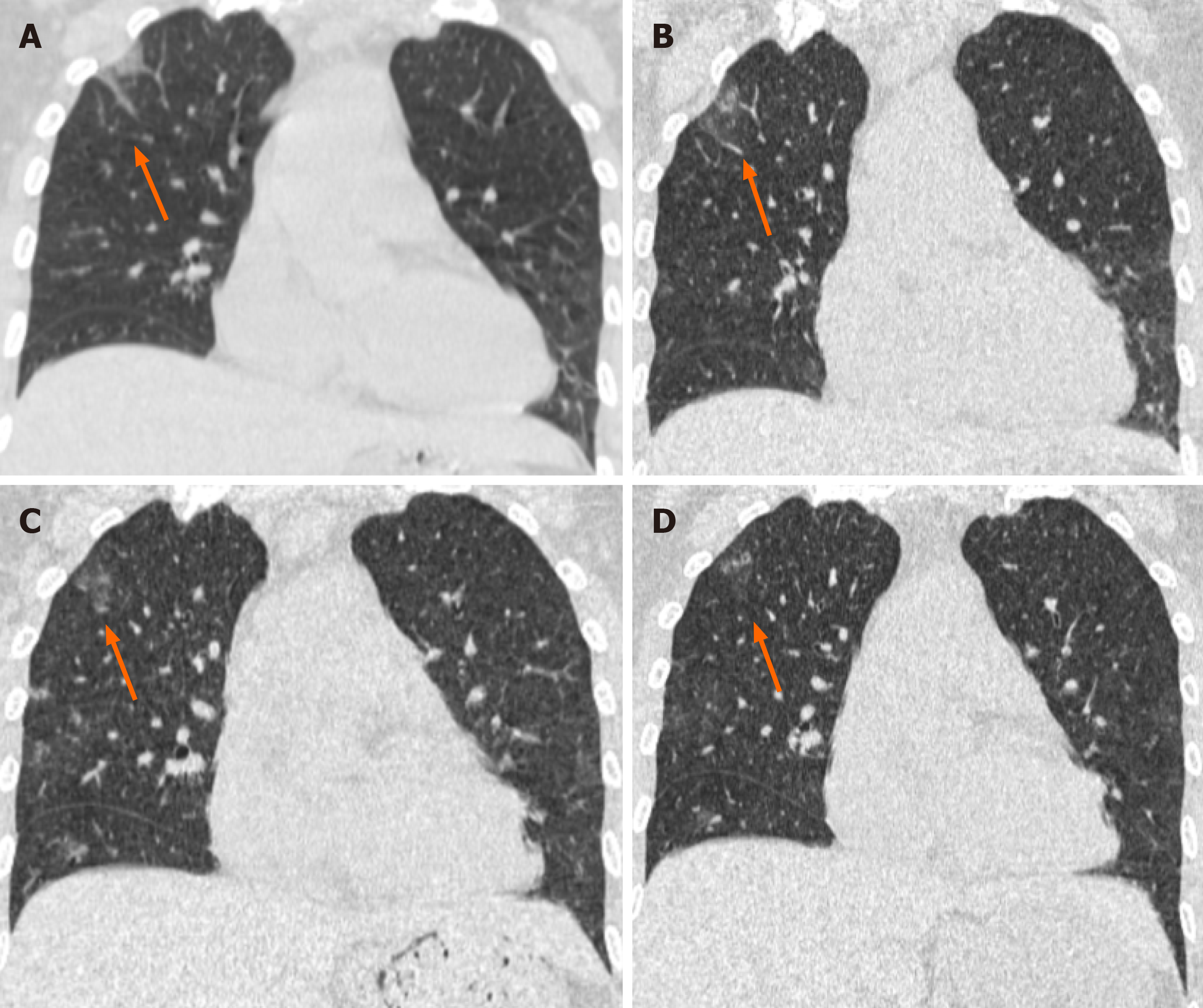
Figure 2 Unenhanced computed tomography images in a 49-year-old woman (coronal imaging).
Computed tomography radiographs show a patchy and striate shadow of slightly high density in the upper right lung (arrows), as the treatment progressed, this asymmetrical lesion was gradually absorbed. A: Day 7 (2020.1.26) after the onset of symptoms; B: Day 12 (2020.1.31) after the onset of symptoms; C: Day 16 (2020.2.4) after the onset of symptoms; D: Day 21 (2020.2.9) after the onset of symptoms.
- Citation: Gao ZA, Gao LB, Chen XJ, Xu Y. Fourty-nine years old woman co-infected with SARS-CoV-2 and Mycoplasma: A case report. World J Clin Cases 2020; 8(23): 6080-6085
- URL: https://www.wjgnet.com/2307-8960/full/v8/i23/6080.htm
- DOI: https://dx.doi.org/10.12998/wjcc.v8.i23.6080


