Copyright
©The Author(s) 2020.
World J Clin Cases. Dec 6, 2020; 8(23): 6026-6035
Published online Dec 6, 2020. doi: 10.12998/wjcc.v8.i23.6026
Published online Dec 6, 2020. doi: 10.12998/wjcc.v8.i23.6026
Figure 1 Computed tomographic presentation of the patient.
A: Mediastinal computed tomographic scan of the chest showed a 2.4 cm × 2.1 cm homogenous well-defined, dense soft tissue lesion in the left lateral inner wall of the trachea (orange arrows); B: Computed tomographic scan with multiplanar reconstruction showed a round lesion in the lower trachea (black arrow); C: A tumor in the inner trachea observed by computed tomographic virtual bronchoscopy (blue arrow).
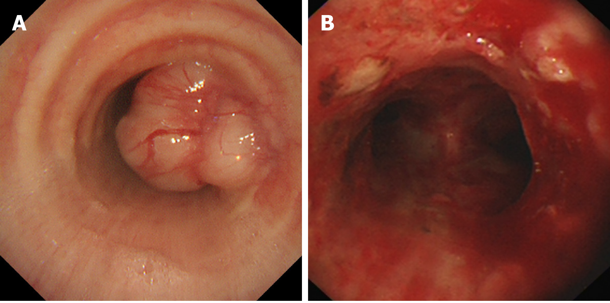
Figure 2 Bronchoscopic findings.
A: A polypoid and vasodilatory mass originated from the right side of the lower trachea; B: After endoscopic resection, the tumor was removed almost completely, and the airway patency was restored.
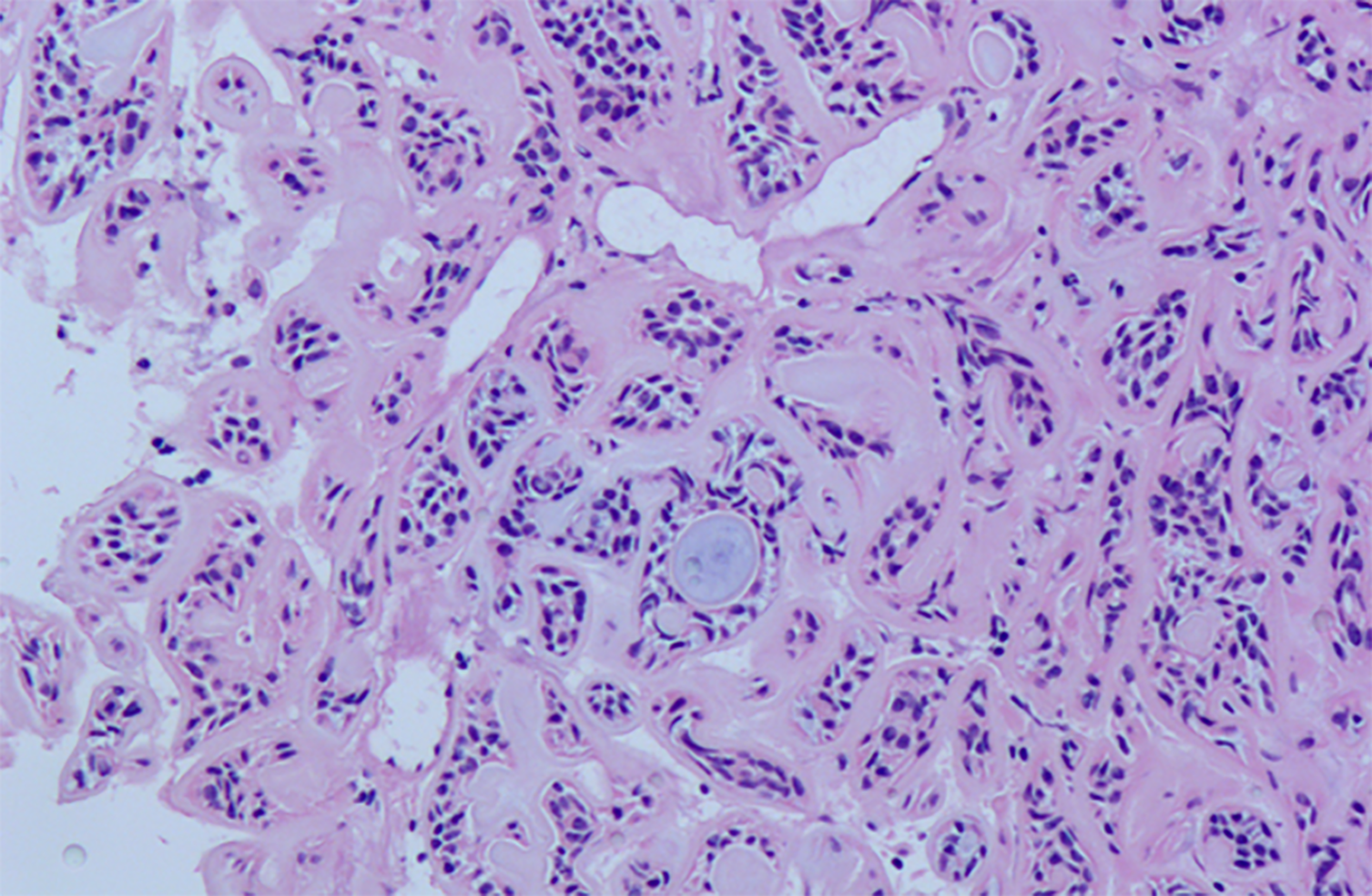
Figure 3 Pathological presentation of the patient.
The tumor was composed of epithelial and myxoid mesenchymal elements and characterized by the presence of ductal structures that appeared to contain double-layered cells in a mucoid or hyaline stroma. No signs of necrosis or mitosis were observed (hematoxylin-eosin staining, × 100).
- Citation: Liao QN, Fang ZK, Chen SB, Fan HZ, Chen LC, Wu XP, He X, Yu HP. Pleomorphic adenoma of the trachea: A case report and review of the literature. World J Clin Cases 2020; 8(23): 6026-6035
- URL: https://www.wjgnet.com/2307-8960/full/v8/i23/6026.htm
- DOI: https://dx.doi.org/10.12998/wjcc.v8.i23.6026


